The landscape of antibody–drug conjugate

Antibody–drug conjugates (ADCs) have become a key therapeutic modality in oncology, spurred by superior clinical profiles compared to standard-of-care chemotherapy across multiple indications. Consequently, revenue from approved ADCs and those in phase III development is forecasted to reach $26 billion in 2028 (Supplementary Fig. 1).

Despite the success of ADCs, long-term growth in their application faces two main challenges. First, the few validated payload mechanisms of action (MoAs) restrict addressable indications. Approved ADC payloads cover three cytotoxic MoAs — anti-mitotic, DNA alkylation and topoisomerase 1 inhibition — that typically require tumour-specific overexpression of the target antigen to ensure sufficient and safe payload delivery. As such, these ADCs primarily target established tumour antigens such as HER2, CD20 and BCMA.

Second, non-specific and insufficient payload delivery narrows the therapeutic window of ADCs. Delivery components for approved ADCs typically include cleavable peptide linkers stochastically conjugated through cysteine reduction to monoclonal antibody carriers. Premature payload release, poor tumour penetration, variable drug-to-antibody ratios and aggregation are common issues.

To explore the impact of next-generation ADC technology on these challenges, we investigated innovation in the ADC clinical pipeline across five design levers — target, payload MoA, antibody, linker and conjugation method — and assessed the likelihood for expanding the addressable indications or widening the therapeutic window of ADCs.

Assessment of the clinical pipeline

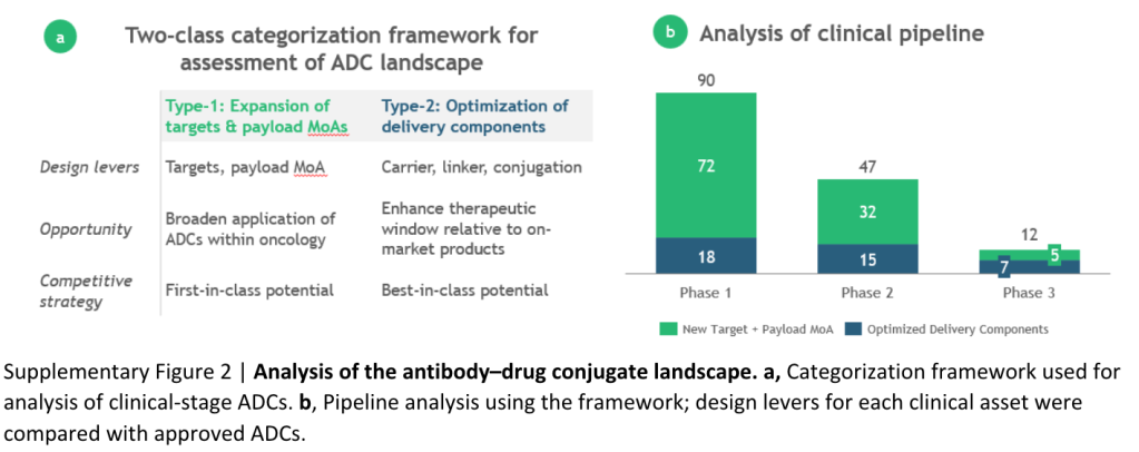
ADC assets in development were categorized into two types based on the potential to overcome the two main challenges (Supplementary Fig. 2a). Type-1 assets have new targets and/or payload MoAs and have first-in-class potential. Type-2 assets leverage established target/payload MoA combinations with novel delivery components to achieve a best-in-class profile.

Daiichi Sankyo’s patritumab deruxtecan, which targets HER3, is an example of a type-1 asset.Recent data from the phase II HERTHENA-Lung01 trial showed an overall response rate (ORR) of 30% in patients with an EGFR mutation and previously treated with an EGFR inhibitor and platinum-based chemotherapy, compared to an estimated real-world ORR of 14% in a similar patient population.

Merck and Kelun Biotech’s SKB264, an example type-2 asset, has the same target and payload MoA as the approved ADC sacituzumab govitecan (Trodelvy; Gilead). SKB264’s differentiated 2-methylsulfonyl pyrimidine linker increases stability in circulation compared to sacituzumab govitecan. An ORR of 40%, with 56% of patients having adverse events (AEs) of grade 3 or higher, was reported for a phase II study  of SKB264 in patients with pre-treated metastatic triple-negative breast cancer. This compares favourably to the 21% ORR and 74% AE rate reported for sacituzumab govitecan  in a similar patient population.

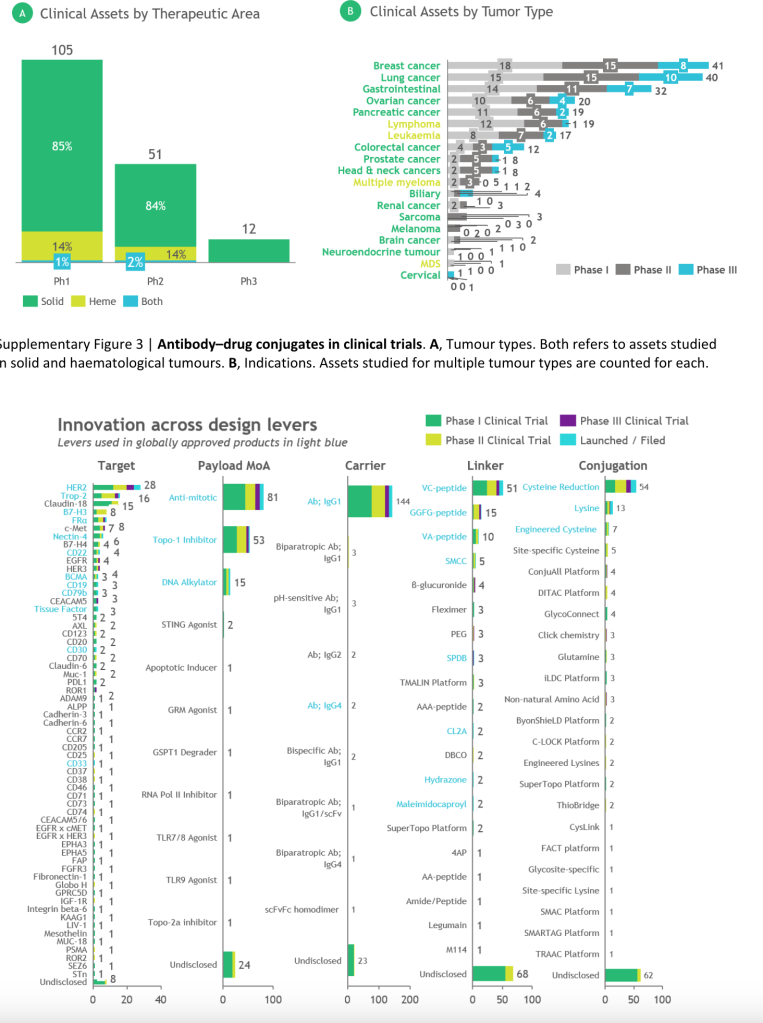
We applied this framework to a database of 168 ADCs in clinical development (Supplementary Fig. 2b). Overall, ~85% of assets address solid tumour indications, with breast and lung cancer the most common (Supplementary Fig. 3). Of the phase III ADCs, ~60% are type-2 assets that leverage established targets and payload MoAs with improved delivery components, which may reflect the recency of the modality’s success and a lower risk tolerance in late-stage development. Greater biological risk is evident in earlier-stage development; ~75% of phase I/II ADCs are type-1 assets with novel combinations of targets and payload mechanisms. Fig. 1 highlights specific components used by assets in development, with the highest concentration in components validated by approved products and a long-tail of single-digit assets with novel components (Supplementary Fig. 4).

Fig. 1 | Assessment of ADCs in clinical development. Innovation across specific design levers used in clinical assets. The top 10 targets and technologies for each lever are shown. Ab, antibody; ADC, antibody–drug conjugate; IgG, immunoglobulin. See Supplementary information for details and an expanded version.

Assessment of next-gen targets

Biological targets are a major innovation area for ADCs, with 61 unique targets under investigation in the clinic. Overall, ~90% of targets are antigens highly expressed on cancer cells, and ~10% of targets are associated with unique characteristics of the tumour microenvironment. For example, Pyxis Oncology’s PYX-201 targets fibronectin, an extracellular protein highly secreted by cancer-associated fibroblasts. ADCs targeting stromal components may prove effective against tumours with  high stromal–tumour ratios such as breast and prostate cancers, and abrogate the evolution of resistance due to the genetic stability of stromal cells.

Assessment of next-gen technology

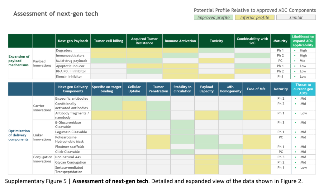
To gauge the potential impact of ADC innovation, we categorized next-gen technology according to our innovation framework and assessed the profile compared with marketed ADCs (Fig. 2). Here, we highlight a subset of next-gen design levers that may be of interest given their novelty and/or promising preclinical and clinical data.

Fig. 2 | Assessment of next-generation ADC technology. Evaluation of next-gen ADC components on potential to either expand ADC applicability or optimize delivery components. Novel biological targets are not considered a technology and therefore not included in the assessment. ADC, antibody–drug conjugate. See Supplementary information for details and an expanded version.

Next-gen payloads. Small-molecule degraders are a promising payload class given their high specificity, picomolar potency and ability to target a broad set of intracellular proteins associated with cancer. Orum Therapeutic’s ORM-5029 delivers a degrader payload selective for GSPT1, a GTPase overexpressed in multiple cancers including gastric, colorectal and breast cancer. Anticancer activity similar to trastuzumab deruxtecan (Enhertu; Daiichi Sankyo) was reported in breast cancer models in preclinical studies. The upcoming phase I data for ORM-5029 will be the first clinical data for antibody–degrader conjugates.

Next-gen carriers. Engineering antibodies with variable antigen binding affinity can reduce off-tissue toxicities and increase tumour-specific exposure. Strategies include shielding Fab domains with peptide masks susceptible to cleavage by proteases overexpressed in tumours, and engineering antibodies with optimized pH-sensitive binding characteristics. Despite past failures, antibody engineering has potential to expand the therapeutic window and treat patients with lower target expression levels. For example, Mythic Therapeutic’s MYTX-011 is designed with lower antigen affinity at endosomal pH to enhance payload escape from endosomes. Increased internalization, cytotoxic activity and in vivo efficacy against c-Met expressing tumour models relative to the parent antibody and a clinical c-Met-targeted ADC was reported in preclinical studies.

Next-gen linkers. Emerging linker technology focuses on controlled payload release independent of endogenous enzyme-mediated cleavage. TagWork’s preclinical ADC TGW101 uses an exogenously administered chemical activator to induce payload release. Superior anti-tumour activity in colorectal and ovarian cancer xenograft models compared to VC-peptide linkers was reported in preclinical studies. Controlled payload release can limit off-tissue toxicity and support further development of assets targeting non-internalizing proteins overexpressed in cancers.

Next-gen conjugation. Incorporation of non-natural amino acids into the antibody carrier facilitates site-specific conjugation through oxime bonds. Ambrx’s ARX788 is a HER2-targeted ADC with a non-cleavable PEG linker attached in a site-specific manner to non-natural amino acids. Phase I data showed anti-tumour activity and improved stability in serum relative to approved HER2-targeted ADCs. Reduction of premature payload release can increase the amount delivered to tumour cells and drive higher response rates.

Conclusion
As ADCs gain traction and new technologies emerge, companies must effectively evaluate emerging platforms, determine investment strategies to maximize expertise and capabilities, and decide whether a first-in-class or best-in-class approach is more attractive. A ‘one-size fits all’ technology is unlikely to emerge in the near-term. Instead, we anticipate companies will build diverse collections of components to enable ‘plug-and-play’ development tailored to specific targets and indications.

内吞体的分离方法

As a functional unit of a living entity, cells are very complicated and consist of exceptionally high number of biomolecules with completely different physical and chemical properties. The complexity of the biomolecules profile is even more intricately woven depending upon their subcellular location. With the limited resolution of the separation and localization technologies, it is imperative to employ fractionation methods for protein profiling and expression analysis.  Even the lesser abundant proteins can be analysed if screened after prefractionation. Sensitive and accurately fractionated samples help in convolution of methods such as mass spectrometry or in-vitro analysis for the detection of proteins at the level of organelles or transportation compartments.

Endosomal organelles are enriched with many proteins that are being transported across the cell for performing vital functions in a compartment-specific manner. Endosomes are membrane-bound vesicular organelles that are crucial for transportation, sorting and synthesis of many macromolecules. Isolation of endosomes provides a lot of information about compartment-specific profiling of the macromolecules.

Compilation of previous studies reflects the spatio-temporal existence of different stages of endosomes such as early endosomes and late endosomes. All these subpopulations vary dramatically in terms of proteomic composition and complexity. Isolation of these endocytic organelles provides a fundamental tool of understanding intracellular signalling events. Over the last few years, various methods were developed to enhance the knowledge about endocytic proteins and their isolation. This blog will discuss briefly about the methods of isolation of endosomes and their further analysis.

Density gradient-based ultracentrifugation

Subcellular fractionation using density gradient based centrifugation provides a great method to isolate endosome organelles and multi-protein complexes.

The overall method has two crucial steps:

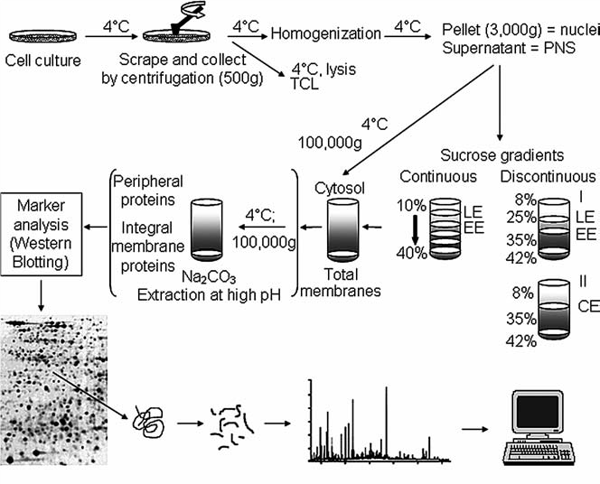
  1. Homogenization: The tissue/cells are homogenized in suitable media to burst open the cell membrane and release the cell organelles and cytoplasmic components into the lysis buffer. The collected cells are briefly centrifuged to remove the cellular debris, unbroken cells, and nuclei. The supernatant obtained at this step consist of cell organelles or membrane bound proteins.
  2. Ultracentrifugation based fractionation: The post-nuclear supernatant obtained after homogenization can be separated into its components using different gradients followed by ultracentrifugation. Discontinuous gradients and step gradient, both of these methods are applicable for the fractionation. The place where a particular membrane or endocytic organelle can be isolated in the gradients is based on the ratio of the lipid content of the endosomes to their protein contents. Heavier cell organelles such as mitochondria and endoplasmic reticulum have high protein content; therefore tend to settle at the bottom of the gradient. Lipid-rich organelles such as endosomes have low density therefore they are isolated from the middle layers or upper layers of the gradient.
    In order to enhance the resolution of the fractionation, the most preferred method for this is using equilibrium separation in a continuous gradient. The post-nuclear supernatant is allowed to equilibrate in a continuous gradient in order to distribute the endosomes of different densities according to their sedimentation quotient. Since the abundance of same density endosome is already low, the only disadvantage of using equilibrium separation is getting a diluted fraction of the endosomes. This drawback can be negated by applying prefractionation approaches such as chromatographic separation, which helps in enriching the sample further.

Endosome_Fractionation_GradientsFigure 1: Schematic outline of fractionation and isolation of endosomal organelles using gradient based ultracentrifugation followed by proteome analysis. Source: Araújo et al., Methods in Molecular Biology. 2008.

Fluorescence-activated organelle sorting (FAOS)

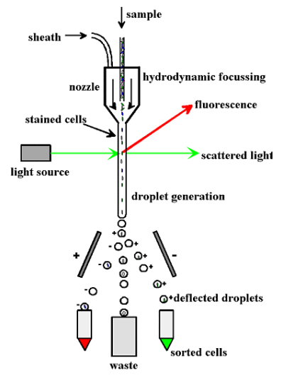
This method utilizes basic principles of flow cytometry to sort and detect the specific endosomes having peculiar light scattering and fluorescent properties. Fluorescent probes, labelled antibodies or fluorescent proteins are used to tag specific organelles. In a way, fractionation is coupled with high speed sorting of the endosomes. The limiting factor in this technique is to decrease the background signal created by the absorption of light by the contents of the sample.

Endosomes_Flow_Cytometry

Figure 2: Diagrammatic representation of FAOS. Source: Stasyk and Huber, Proteomics, 2005. 

Free Low electrophoresis (FFE) and Flow Field-Flow Fractionation

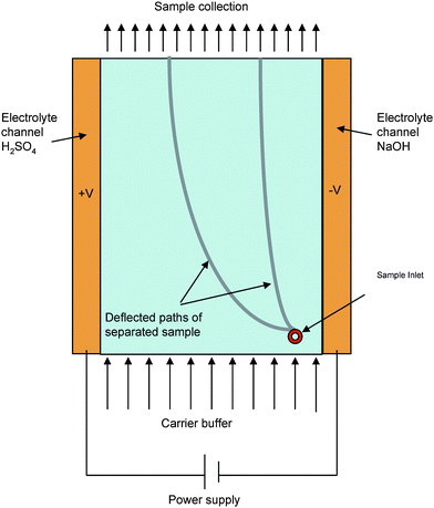
This method harnesses the property of differential mobilities of different organelles based on their membrane properties when exposed to an electric field followed by collection of the sample at different time points. Principally, the sample is allowed to move in a laminar flow manner and then a suitable electric field is applied perpendicular to the flow. The resultant potential causes the displacement of organelles along the electric field. The final displacement and mobility of the organelles is decided by the surface biomolecules and the net charge on them. This is a great tool to isolate an enriched fraction of the organelles especially the small ones such as endosomes. Each fraction can be used for further analysis based on the organelle specific biomarkers.

Free_Flow_Electrophoresis

Figure 3: Schematic representation of a free flow electrophoresis. Source: Ho S et al, Journal of Materials Chemistry, 2009.

Immunoisolation of endosomes

This method relies on binding separation of organelles using their antigenic properties rather than using their physical properties, unlike other isolation methods. The basic principle is closely similar to antibodies and antigen binding as any other immunological method. However, the parameters of choosing antigens, antibodies and the support on which it is growing vary substantially.

  1. Antigens: Immunoisolation works on a one to one ratio which suggests a single epitope is enough for separating a single vesicle. Therefore, the epitope that should be chosen must be exclusively present on the desired endosomal compartment. Since, this method does not consider the variation in physical properties such as density of the antigen; therefore, this method is not suitable for differential immunoisolation. Care should be taken about the accessibility of the selected epitope. It should be easily accessible to the antibody which is immobilized on a solid surface. It is always suggested to select the epitope which has higher abundance to increase the efficiency of binding.
  2. Antibodies: Antibodies are held on a solid support in this method, therefore two different types of antibodies are used for an efficient binding.
    1. Linker antibody- it is used to enhance the range of binding and flexibility of the specific antibody.  It couples the solid surface beads with the specific antibody to allow endosomal binding. Mostly commonly a generic anti-Fc antibody is used as a linker antibody.
    2. Specific antibody: These antibodies are raised against the epitope present on the surface of the endosomes that are required to be isolated. It is suggested to use an affinity purified polyclonal antibody. However, the binding of antibody with the endosomes require optimisation and calibration.
  3. Solid Support/matrix:Binding of the linker antibody on a solid surface is crucial. It is also imperative to choose a solid matrix carefully.
  • Flexibility: An ideal matrix should be flexible enough to bind with the antibody without a positional disorientation and conformational hindrance.
  • Sedimentation: Matrix should be able to aggregate and sediment easily at a very low speed like 3000 x g at which the endosomes do not co-sediment. It should not be cross linking with the endosomes as well so that both of these can be easily separated after binding.

The most common modification of immunoisolation is the use of Magnetic beads. This approach is beneficial in being rapid, gentle and efficient. It does not require high speed centrifugation or density gradient based fractionation for separation of endosomes. It is also a useful method to isolate differential densities of endosomes with greater efficiency and purity.

Immunoisolation

Figure 4: Schematic representation of magnetic beads based immunoisolation. Source: Lordachescu A et al., Royal Society of Chemistry Advances, 2018

 Magnetic beads can be used in two ways:

  • Direct Binding: The magnetic beads are incubated with the antibody first for a short duration followed by a longer incubation with the crude sample containing endosomes. The targeted endosomes attached with the beads can then be separated by using a magnet and few washing steps.
  • Indirect Binding: In this protocol, the antigen and the antibodies are allowed to bind first. Magnetic beads are added with the antigen/ligand mix later on and allowed to bind for some time. The extraction and purification method is similar as that of direct binding technique.

Direct binding technique is recommended when the antigen is easy to access. When the targeted antigen is difficult to access, the indirect binding technique is preferred.

Points to be considered during endosomal isolation:

  1. Nuclear rupture while homogenisation releases DNA resulting into increased viscosity of the solution. Therefore, enough detergent has to be added to maintain the consistency of the homogenate.
  2. Cross contamination of the samples should be avoided from the standards.
  3. Endosomes are very fragile under ionic conditions. Therefore, the homogenisation of the tissue should be very mechanical and mild in order to avoid their rupture.
  4. While making gradient in the density-gradient fractionation, air bubbles should be avoided as they interfere with the continuity of the gradient.
  5.  The layers after ultracentrifugation should be collected carefully so as to obtain a pure population of the organelles.
  6.  Endosomal population obtained after the method should be checked for purity using endosomal markers.

来源: https://info.gbiosciences.com/blog/four-methods-for-endosomal-isolation

Kill switch for CAR-T

How researchers are addressing risks and opportunities in this groundbreaking area of oncology research

Last August, in a milestone decades in the making, Kymriah became the first cell therapy to be approved by the US Food and Drug Administration (FDA). The approval was based on a groundbreaking clinical study that showed the drug could wipe out the blood cancer in people who had already gone through many unsuccessful rounds of traditional therapy. Just three months after receiving Kymriah, 83 percent of these patients were in complete remission.

Kymriah was the first success story for a new kind of treatment called chimeric antigen receptor T-cell therapy, or CAR-T therapy. These remarkable results set off a frenzy in the biotech world: The same month that Kymriah was approved, Gilead Sciences agreed to pay $11.9 billion for Kite Pharma, which subsequently received FDA approval for its own CAR-T therapy, Yescarta. Later, Celgene paid $9 billion to acquire Juno Therapeutics, a biopharmaceutical company founded in 2013 through a collaboration of the Fred Hutchinson Cancer Research Center, Memorial Sloan-Kettering Cancer Center and pediatrics partner Seattle Children’s Research Institute, that doesn’t yet have an FDA-approved therapy.

Unlike conventional drugs that are manufactured in bulk, Kymriah and Yescarta must be made new for every patient. The process starts by removing a person’s T cells, inserting a gene for a synthetic protein called a chimeric antigen receptor, then reinfusing it into the patient to spot and destroy tumors.

The price of remission

Despite all of the excitement generated by these results, there are still significant limitations to CAR-T cell technology. Kymriah and Yescarta treat only people with rare blood cancers. And personalizing medicine comes at a price: A one-time injection of Kymriah costs $475,000. Add the related hospital care and the overall cost of CAR-T therapy can approach $1 million. Health insurers are still grappling with how to pay for that. Although it’s early in Kymriah’s adoption, the therapy brought in only $12 million in sales last quarter.

One of the other issues with CAR-T cells is that they can produce severe systemic inflammation (cytokine release syndrome or CRS) as well as neurologic toxicities, which can be difficult to treat. These toxicities have been responsible for the death of more than a dozen people in clinical trials. Tocilizumab, an anti-IL6 antibody, which was approved for the treatment of several autoimmune diseases has been successful at reducing some of the symptoms of CRS, and has made CAR-T therapy somewhat safer, but it has only modest impact on overall neurological dysfunction. Because of the price, as well as risk of toxicities, CAR-T cell therapy is reserved as a last resort for patients with no other options.

Successful applications of CAR-T therapy have also been more elusive for more common solid tumors, such as lung, colorectal and breast cancer. Solid tumors release a slew of molecules to suppress the immune system and shield themselves from T-cell attack.

The lack of CAR-T efficacy in solid tumors can be due to the environmental barriers that exist (e.g., endothelial cell dysfunction, extracellular matrix barriers, or lack of nutrients,) which prevent migration and infiltration to physically access the tumor. Many tumors also have chronic inflammation with immunosuppressive molecules that express immune checkpoint inhibitor ligands, inducing strong immunosuppression within the tumor microenvironment.

To improve CAR-T function in solid tumors, it is important to focus on improving tumor targeting; defining optimal CAR structures and signaling domains; and synergizing CAR-T cells with the pro-immune and anti-tumor effects of oncolytic viruses, checkpoint inhibitors, and cytokine delivery.

Suicide cells and GoCAR-Ts

There are a growing number of biotech companies devising control systems to make new CAR-T therapies simultaneously safer and more powerful.

  1. An emergency “kill” switch to the CAR-T cells to address the onset of CRS. Several firms are working on such solutions, including Houston-based Bellicum Pharmaceuticals, which has engineered a safety switch using two engineered proteins that dimerize when exposed to a small-molecule drug called rimiducid. Rimiducid activates a protein called caspase-9, which initiates the process of CAR-T-cell suicide.
  2. Antibody-based safety switch to induce the suicide of CAR-T cells. The French company Cellectis tags its CAR-T cells with a protein that the cancer antibody rituximab—marketed as Rituxan – can bind. A rituximab injection should cause a patient’s normal immune cells to target and kill the CAR-T cells. Cellectis is working on new versions of CAR-T cells that can be activated or deactivated with small molecules.
  3. Default state to off. Bellicum has a second CAR-T design that features a default state set to off, engineered to turn on only when the CAR binds a cancer antigen and an activating small molecule drug simultaneously. These so-called GoCAR-Ts should allow doctors to increase T-cell activity and proliferation by raising the dose of that drug, which is rimiducid. Bellicum rewired its GoCAR-Ts to be activated by rimiducid, rather than killed by it, as in the suicide switch-containing cells. This design could allow doctors to scale back CAR-T activity by reducing or halting administration of rimiducid. But the main goal for GoCAR-Ts is to drive the therapy into solid tumors.
  4. Extended life CAR-T: Bellicum is also attempting to prolong the effective life of the CAR-T cells by using rimiducid to repeatedly pump the gas on the GoCAR-T cells as they search for solid tumors. Interleukin 12 (IL-12) is a potent cytokine known to counteract the tumor’s immunosuppressive environment. It is too potent to be taken as a drug, but letting CAR-T cells produce their own IL-12 could be a safer way to overcome a tumor’s suppressive barriers. Boston-based Ziopharm Oncology is one of several other companies developing CAR T cells that produce either IL-15 or IL-12.

https://www.iqvia.com/blogs/2019/01/a-suicide-switch-for-car-t

https://www.mdpi.com/2073-4409/11/12/1910

Choosing the right tool for designing guide RNAs

https://www.takarabio.com/learning-centers/gene-function/gene-editing/gene-editing-tools-and-information/sgrna-design-tools

The first step of CRISPR/Cas9 gene editing is designing a single guide RNA (sgRNA) to target your gene of interest. Because sgRNAs are solely responsible for recruiting Cas9 to specific genomic loci, optimal sgRNA design is critical for successful gene editing experiments. There are many web-based tools available for sgRNA design, each of which has different features and advantages. The information provided here will help you choose the best tool for your specific research objective.

Several web-based tools available

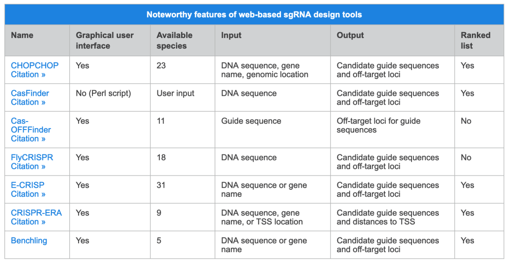
Web-based sgRNA design tools typically require that users input a DNA sequence, genomic location, or gene name for each gene of interest, and indicate a species. An algorithm specific to each tool outputs a list of candidate guide sequences with corresponding predicted off-target sites for each input (Wu et al. 2014). Most tools aim to provide guide sequences that minimize the likelihood of off-target effects, but the methods they employ vary. For example, Chop Chop uses empirical data from multiple recent publications (e.g. Doench et al. 2016) to calculate efficiency scores. Alternatively, CasFinder (Aach et al. 2014) and E-CRISP (Heigwer et al. 2014) incorporate specific user-defined penalties based on the number and position of mismatches relative to the guide sequence in order to rank the potential for off-target effects.

Tools for specific applications

Some sgRNA design tools have been developed for specific applications. CRISPR-ERA (Liu et al. 2015) is the only currently available tool that designs sgRNAs specifically for gene repression or activation, while FlyCRISPR (Gratzet al. 2013) focuses on applications in fly, beetle, and worm species, including the popular model organisms Drosophila melanogaster and Caenorhabditis elegans. Presently, the design tool featured on the Benchling website is the only one that can generate candidate sgRNAs that are compatible with alternative nucleases such as Staphylococcus aureus Cas9 (Ran et al. 2015) and Cpf1 (Zetsche et al. 2015). Given the uniqueness of each tool, we recommend that you use multiple approaches during the sgRNA design process and choose guide sequences that are consistently predicted to perform well.

A selection of freely available tools

The table below provides a list of web-based tools for sgRNA design. For simplicity, we have only included those that are free and do not require a subscription. For each tool, we have indicated whether there is a convenient graphical user interface or if the user has to download a script. If you would prefer to design sgRNAs manually, please visit Choosing a target sequence for CRISPR/Cas9 gene editing to learn more.

神经退行性疾病的机制

https://ars.els-cdn.com/content/image/1-s2.0-S0092867422015756-gr2_lrg.jpg

病理性蛋白聚集

蛋白聚集这一特征经常被作为疾病诊断和分类的依据,相关的疾病包括阿尔兹海默(Alzheimer disease, AD),帕金森(Parkinson disease, PD),原发性陶氏病(primary tauopathies),额颞痴呆(frontotemporal dementia, FTD),肌萎缩性脊髓侧索硬化症(amyotrophic lateral sclerosis, ALS)等。对于很多NDDs而言,蛋白聚集都发生在脑区,与临床预后相关,提示其可能发挥病理作用。遗传学角度的解析发现NDDs中有很多突变,这会导致有毒功能的积累,正常功能的丧失,另外还有因为朊蛋白的错误折叠,从而促进病理过程。相关的生物标志物有Aβ42/Aβ40,磷酸化的tau等。但需要注意的是,蛋白聚集并不总是与疾病进展完美匹配,还有许多其他的机制造成神经毒性。另外还有一些非蛋白致病性的NDDs,例如创伤性脑损伤(traumatic brain injury, TBI),慢性创伤性脑部病变(chronic traumatic encephalopathy, CTE),中风等。

突触和神经元网络功能障碍
NDDs的症状典型地反映了特定神经网络的紊乱,神经元网络功能的发挥依赖于突触功能的精准调控,而突触的损伤和毒性似乎是在神经元损伤前就出现的。突触功能受线粒体功能调控,需要精准的能量调节以维持钙稳态和离子平衡,星形胶质细胞和小胶质细胞也在能量,神经递质稳态,突触消除和稳定中发挥着重要作用。研究表明多种NDDs与突触功能障碍相关,例如在AD,PD,FTD中,突触功能障碍是早期事件,这一事件同时得到了成像的验证。由于涉及到神经元网络功能异常,它会同时与多个NDD特征相关。

蛋白质稳态异常
蛋白质稳态的维持依赖于泛素-蛋白酶体系统(UPS)和自噬-溶酶体途径(ALP)这两种主要的细胞机制,NDDs中泛素化聚集蛋白的积累提示蛋白质稳态发生了异常。UPS两个组分UBQLN2和VCP的突变与ALS/FTD相关,NDDs中聚集的一些蛋白也会损害UPS的功能。类似地,脑组织特异的自噬失活会导致NDDs和聚集蛋白积累,PD相关的基因和蛋白都在自噬和溶酶体功能中发挥重要作用,它们能够造成ALP和突触功能异常,影响细胞-细胞间蛋白运输和神经细胞死亡。

细胞骨架异常
神经元的细胞骨架主要有3个聚合结构:基于微管蛋白的微管,中间纤维,以肌动蛋白为基础的微丝,三者之间互相关联但蛋白组分和直径不同。这些结构是神经元建造维持,组织运输的基础,支撑着能量稳态和突触功能。NDDs中神经元细胞骨架改变,传递信息,交换物质的能力丧失,主要涉及细胞骨架相关基因的突变如中间纤维基因的突变,轴突运输机器的突变,蛋白的异常磷酸化和失调,神经丝和肌动蛋白的异常聚集等。细胞骨架的破坏与其他NDD标志间也存在着互作,尤其是突触维持的丧失和能量代谢的改变,RNA运输,蛋白质聚集和自噬,以及神经元死亡。

能量稳态改变
多种NDDs中都呈现出能量代谢缺陷。ATP是大脑能量代谢的关键分子,其由葡萄糖或乳酸代谢产生,这两个能量底物能够通过星形胶质细胞直接从血流传到神经元。以往的研究表明,低ATP利用率与线粒体功能障碍等都能诱发神经元功能失调甚至是细胞死亡。神经退行性疾病中风就是一个很好的例子,兴奋毒性和能量耗竭共同促进疾病进展。此外,糖酵解,脂代谢,线粒体代谢中的酶的遗传缺陷也会导致神经系统出现故障。类似的机制还有线粒体DNA的突变等,而线粒体的损伤又会进一步影响Ca2+稳态,从而影响与之相关的多种生物学过程,由此与其他NDD标志物也紧密相连。

DNA和RNA缺陷
DNA层面的改变能够驱动突变,染色质重排等多种不利分子事件,RNA代谢和稳态的缺陷会导致RNA转运等生物过程异常。DNA损伤的危害在几种罕见遗传性疾病中表现得尤为明显,它会削弱响应和清除基因组应激的能力,氧化DNA损伤更被提出可作为AD模型和PD样病理的风险因子。此外,NDDs中常见的蛋白也参与不同的DNA损伤应答,DNA损伤与DNA损伤感知器PARP1的超活化和线粒体功能失调有关。部分NDDs患者呈现出RNA调节异常,主要是代谢改变,多系统的蛋白质病变等,从而影响RNA稳态。

炎症
神经炎症包括小胶质细胞增生和星形胶质细胞增生,前者存在于所有NDDs中,小胶质细胞负责大脑感知,管家和防御功能,NDDs中聚集蛋白等危险信号会激活小胶质细胞,调节细胞因子,ROS的产生,与星形胶质细胞的互作等,与其功能相关的很多基因都在AD中被鉴定出来。与小胶质细胞联系紧密的星形胶质细胞也对神经元的健康和功能十分重要,尤其是在谷氨酸稳态和三突触网络中,其与小胶质细胞间的紧密互作可能还会共同促进NDD进展。

神经细胞死亡
神经元一些固有特性使得其特别容易受到细胞死亡的影响。首先,有丝分裂后细胞意味着其逐渐积累年龄相关的损伤以及无法复制更新;其次,由于需要供给突触功能和ROS产生,对能量要求极高;延展的轴突树突意味着运输和结构组织需要远距离实现;依赖胶质细胞来维持,产能和防御。前面提到的NDD相关标志特征都与神经元损伤有关,这最终会导致脑容量损失和神经细胞内蛋白的释放。关于神经元死亡有两个比较公认的机制:内在外在的凋亡和细胞坏死。其他机制包括坏死性凋亡,铁死亡等也发挥作用,不同机制间存在crosstalk。与细胞死亡相关的分子层面途径包括线粒体渗透性过渡孔(mPTP)的打开,突触前和生长树突电压依赖的Ca2+通道的激活,不依赖caspase的染色质凝结等。此外,非细胞自主性死亡也导致神经元丢失。

https://www.sciencedirect.com/science/article/pii/S0092867422015756

genome-editing for cardiomyopathy

Genome editing has progressed rapidly from discovery to clinical development, while preclinical studies continue to refine the approaches. Now, two papers in Nature Medicine showcase the potential of two such strategies — base editing and CRISPR–Cas9 — for prevention of hypertrophic cardiomyopathy (HCM) in mouse models. The studies also highlight different advantages and challenges for each genome-editing strategy.

HCM is caused by mutations in cardiac sarcomeric genes that lead to thickening of the heart muscle and can cause heart failure and sudden cardiac death. A dominant-negative pathogenic variant in the sarcomeric protein β-myosin, c.1208G>A, is a well-studied cause of severe HCM.

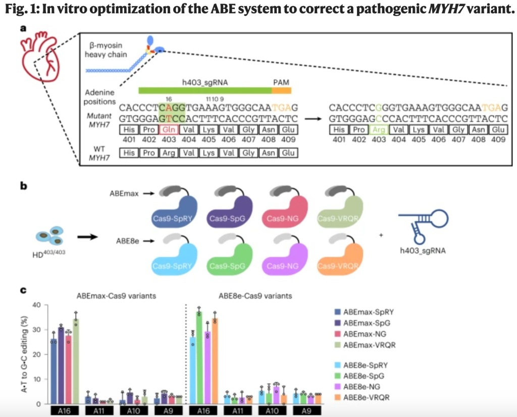
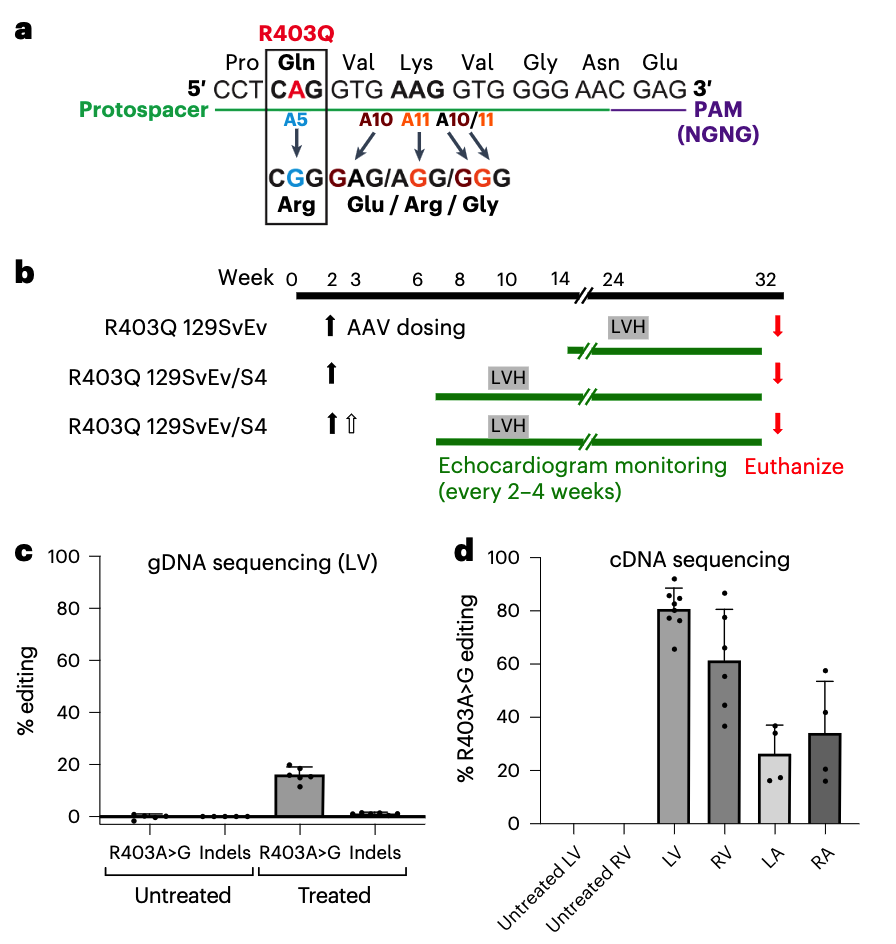
In one of the studies, Chai et al. set out to correct the c.1208G>A mutation using a base editor — a fusion protein consisting of a modified Cas9 nickase and a deaminase enzyme that converts one DNA base into another, at a site determined by a guide RNA (gRNA) sequence.

The authors used human induced pluripotent stem cells (iPSCs) to screen various adenine base editors (ABEs) with different editing efficiency and specificity. They opted for an ABE with a narrow editing window. Although this had a relatively low efficiency (34%), the risk of bystander edits — whereby the base editor modifies other adenine residues close to the target adenine — was also low.

In cardiomyocytes generated from patient-derived iPSCs heterozygous for the c.1208G>A mutation, treatment with the base editor reduced contractile force generation and ATP consumption back to normal levels. The researchers detected minimal bystander editing with little to no off-target editing at distant DNA sites.

Next, Chai et al. generated a mouse model that carried human sequences encoding the β-myosin pathogenic variant. Owing to the large size of the base editor enzyme, they delivered it over two adeno-associated virus 9 (AAV9) vectors, and used a troponin T promoter to target expression to cardiomyocytes.

Mice received intrathoracic injection of the AAV9-vectored base editor immediately after birth, prior to development of HCM. Compared with untreated mice, at 8–16 weeks ABE-treated mice showed reduced features of HCM, such as ventricular wall thickening, and had similar echocardiographic readouts to wild-type mice.

The investigators detected a 32% editing efficiency of the target pathogenic adenine in cardiomyocytes, with no bystander editing. Low-level editing in off-target tissue such as the liver was detected.

In the other study, Reichart et al. tested both an ABE and a CRISPR–Cas9 nuclease that would respectively correct or silence the β-myosin mutation.

They selected an ABE with high editing efficiency, which corrected the mutation in >70% of left ventricular cardiomyocytes. Treatment of mouse models at 10–13 days of age prevented onset of HCM cardiac morphology and dysfunction for 32 weeks. However, bystander edits occurred at a rate of 3–5%, and a low but significant rate of off-target editing was detected.

Reichart et al. also designed a CRISPR–Cas9 nuclease system to selectively inactivate the c.1208G>A mutation in cardiomyocytes. Here, the gRNA directed Cas9 nuclease to make double-stranded breaks (DSBs) and generate indels in the target gene. The smaller size of Cas9 compared with ABEs enables the therapeutic to be packaged in a single AAV. And although this approach cannot correct a mutation, it is more amenable to application across different mutations.

Intrathoracic injection of the CRISPR–Cas9 prevented HCM onset in mouse models. However, high-dose treatment was associated with impaired cardiac function resulting from editing of the wild-type allele, suggesting a narrow therapeutic window.

Further studies and emerging clinical data will assist researchers in balancing editing efficiency with safety and other parameters to select appropriate genome-editing tools.

1- Chai, A. C. et al. Base editing correction of hypertrophic cardiomyopathy in human cardiomyocytes and humanized mice. Nat. Med. 29, 401–411 (2023)

2- Reichart, D. et al. Efficient in vivo genome editing prevents hypertrophic cardiomyopathy in mice. Nat. Med. 29, 412–421 (2023)

Inhibiting cap snatching

To prime their own transcription, influenza A virus (IAV) and influenza B virus (IBV) use ‘cap snatching’, in which the 5′ end of fully capped host RNA is removed and attached to viral mRNAs. Deficiency in host RNA methyltransferase MTR1 — which mediates a 2′-O-methylation step required for host cap maturation — has previously been shown to enhance antiviral interferon responses to the IAV, reduce its cap-snatching efficacy and impair replication. MTR1 may therefore represent a potential target for anti-influenza drugs. Here, Tsukamoto et al. demonstrate in a human cell line that MTR1 is essential for the initiation of replication of both IAV and IBV. An in silico screen of 5,597 compounds, followed by molecular docking studies using the crystal structure of MTR1, identified the adenosine analogue, tubercidin — a natural product from Streptomyces — as an inhibitor of MTR1. Evaluation of 115 tubercidin-related compounds in antiviral drug assays revealed trifluoromethyl tubercidin (TFMT) to be the most effective non-toxic compound. TFMT inhibited replication of various strains of IAV and IBV in human bronchial epithelial cells, human lung explants ex vivo, and mice, without signs of toxicity. In vitro mechanistic studies confirmed TFMT to inhibit IAV cap snatching, acting synergistically with antivirals baloxavir marboxil and oseltamivir.

Tsukamoto, Y. et al. Inhibition of cellular RNA methyltransferase abrogates influenza virus capping and replication. Science 379, 586–591 (2023)

Peptide barcodes meet drug discovery

The success or failure of small-molecule drug discovery efforts strongly depends on the “hit-finding” approaches that are applied at the inception of the drug discovery program (1). High-throughput screening of compound collections is still the main strategy (2), but several other approaches have shown promise. These include screening virtual libraries using three-dimensional protein structure or ligand information (3), de novo design of ligands (4), screening fragment (very small molecule) libraries (5), screening (cyclic) peptide libraries (6), repurposing existing compounds, and screening DNA-encoded libraries (DELs) (7). On page 939 of this issue, Rössler et al. (8) reveal a new hit-finding method that uses peptide-encoded libraries (PELs), which are similar to DELs.

In PELs, solid-phase peptide and small-molecule syntheses are used to readily generate large libraries of bifunctional molecules that each consist of a peptide tethered to a small molecule through a cleavable linker. After cleavage from the solid phase, these libraries are incubated with an immobilized therapeutic protein of interest for affinity selection. To identify those molecules that bind to the target protein, the peptide is cleaved from the bifunctional molecule and sequenced using mass spectrometry technologies that are normally applied in proteomics research (such as nanoscale liquid chromatography. tandem mass spectrometry). On the basis of the sequence of the peptide, the chemical structure of the smallmolecule ligand can be identified. This is because the single amino acids that are used for synthesis of the peptide directly encode the corresponding chemical building blocks that are used to synthesize the small molecules.

The advantages of PEL technology over DELs are manifold. Most notably, a PEL supports harsher and more diverse chemical reactions, including metal-catalyzed reactions and reactions that require strong acidic or basic conditions. This breadth enables the synthesis of a wider scope of drug-like molecules. Another advantage is the application of solid-phase synthesis for peptides and small molecules, which allows the use of excess reactants. This, in turn, supports a higher yield and purity of the final small molecules, which is expected to substantially improve the quality of the libraries. Changing the tagging moiety from four DNA bases to a peptide that contains 16 different amino acids enables a higher information capacity. Thus, in theory, even larger libraries of small molecules could be synthesized and encoded. If an eight-digit encoding string is used, then there are 16 amino acids (hexadecimal system) that can generate 4.3 billion possible codes. By contrast, there are only 56,535 possible codes using the four bases of DNA.

It is thought that the DNA tag of the DELs could interfere with targets that are per se DNA-binding, such as transcription factors or RNA. By contrast, libraries with peptide tags would potentially be better suited for screening against such targets because the amino acids used for the peptide synthesis are less likely to bind to those targets. To confirm that a hit identified in a DEL screen can actually bind to a target, the hit compound is synthesized without a DNA tag and then tested for its effect on biological activity. This can be tedious because, during DEL library synthesis, not every chemical reaction is successful. Occasionally, reaction byproducts are the biologically active compounds, and it takes several investigations to determine this. The hit resynthesis that stems from a PEL can still be performed by solid-phase synthesis using the same conditions that were used to construct the library. This allows a more rapid synthesis and makes the identification of potential by-products easier.

Some challenges need to be overcome to fully exploit PEL technology. Peptide concentrations must be present in at least a 10 fM range to be detected by mass spectrometry. This affects the size of a PEL because, in contrast to a DNA tag, the peptide tag cannot be amplified. The screens of Rössler et al. were performed at ∼1 nM concentration for each peptide-tagged compound. This means that a 100,000-membered library could be screened at a 100 µM library concentration. Because the peptide tags used by Rössler et al. were mostly hydrophobic, there is a certain risk of solubility problems and unspecific peptide aggregation of the library members, which could interfere with binding to a putative target and lead to screening artifacts.

The libraries generated by Rössler et al. were screened against the targets human carbonic anhydrase IX, the epigenetic reader bromodomain-containing protein 4 (BRD4), and the E3 ubiquitin ligase mouse double minute 2 homolog (MDM2). In all cases, several hits were identified that could serve as interesting starting points for further improvements of their potency and properties. PELs could potentially be enhanced by exploiting a wealth of already-established solid-phase organic chemistry reactions to generate new druglike molecules in a chemical space that is not accessible by the DEL technology. Such libraries would be of high interest to drug discovery groups for screening against therapeutic protein targets for which no small-molecule ligands are yet known.

REF ERENCES AND NOTES
1. D. G. Brown, J. Boström, J. Med. Chem. 61, 9442 (2018).
2. P. S. Dragovich, W. Haap, M. M. Mulvihill, J.-M. Plancher, A. F. Stepan, J. Med. Chem. 65, 3606 (2022).
3. F. Gentile et al., Nat. Protoc. 17, 672 (2022).
4. M. Skalic, J. Jiménez, D. Sabbadin, G. De Fabritiis, J. Chem. Inf. Model. 59, 1205 (2019).
5. D. A. Erlanson, S. W. Fesik, R. E. Hubbard, W. Jahnke, H. Jhoti, Nat. Rev. Drug Discov. 15, 605 (2016).
6. C. Sohrabi, A. Foster, A. Tavassoli, Nat. Rev. Chem. 4, 90 (2020).
7. R. A. Goodnow Jr., C. E. Dumelin, A. D. Keefe, Nat. Rev. Drug Discov. 16, 131 (2017).
8. S. L. Rössler, N. M. Grob, S. L. Buchwald, B. L. Pentelute, Science 379, 939 (2023)

入职后,该储备哪些知识和技巧?

从一个大药企出来后,脑海里残存的记忆已经淡去。但是资源不能带出去,如何把每一段经历融入自己的生涯呢?

第一,入职后,总结面试成功和失败的经验。分析自己的哪些方向受市场欢迎,哪些是自己的弱项,需要在今后的工作中加以提升和改进。

第二, 入职后,开会时勤记笔记。电子笔记的泛滥让人们怠于手写笔记,一切都是复制粘贴。而电子化的媒介是不能携带出去的。所以及时笔录备份脑海里的记忆,是非常重要的。

第三,在工作过程中,要时常回顾面试中问及的问题,尤其是behavior中的问题。只有一点点积累,才能在面试中轻松准备。例如:https://www.indeed.com/career-advice/interviewing/director-interview-questions。https://passmyinterview.com/director-interview/ https://posts.careerengine.us/p/5c2d66f349664b22db8a2b5b

第四,

Neutralizing antibodies for AAV vectors: The strange case of AAV5

The recent approval by the U.S. Food and Drug Administration of gene therapies for eye and muscle disorders and the growing number of clinical trials with adeno-associated virus (AAV)–derived vectors clearly indicates the maturity of this method of gene replacement for the use in humans. The large clinical experience in liver gene transfer with AAV mostly derives from clinical trials for the treatment of two coagulation disorders, hemophilia A and B. Data from different trials strongly support the safety and the efficacy of the approach.

One major limitation of the systemic administration of AAV is the presence of preexisting neutralizing antibodies against the vector. Indeed, seropositivity for AAV is among the exclusion criteria in most of the AAV gene therapy trials. Early studies indicated that very low titers of neutralizing antibodies in circulation prevented vector entry and resulted in reduced liver transduction. So far, patients injected with AAV vectors had little to none anti-AAV neutralizing titers. This was true until the results of the clinical trial sponsored by uniQure for the treatment of hemophilia B with an AAV5 vector expressing human coagulation factor IX (hFIX) were public.

The assay used to measure preexisting neutralizing antibodies in the patients of the trial was based on green fluorescent protein (GFP) as a reporter and had a limited sensitivity. By using a more sensitive assay, based on luciferase as reporter, Majowicz et al. demonstrated that three of the patients included in the clinical trial were seropositive for AAV5 with titers that have been associated with in vivo neutralization of liver transduction in preclinical animal models. Importantly, in the patient who had the highest neutralizing titers, the expression of hFIX was similar, if not superior, to that of patients of the same dose cohort. This suggests that the serotype used in the trial, for some reason, was less sensitive to antibody neutralization. To support the clinical data, they performed an in vivo neutralization assay in nonhuman primates (NHPs). In this experiment, they tested four increasing doses of vector in a range that cover the doses of AAV normally administered in the clinic. The NHPs dosed were all seropositives for anti-AAV5 neutralizing antibodies with titers spanning from low (1/57) to relatively high (1/1030). Interestingly, they did not see the expected inverse correlation between the neutralizing titers and the transduction efficacy as measured by hFIX protein secreted in circulation.

One important caveat in the interpretation of the results of this study is the absence of standardized methods to measure vector and neutralizing antibody titers. This represents a major limitation in the field of gene therapy that hampers the comparison of data between the different laboratories and the exact reproduction of the data. Despite this, in 2018, Biomarin announced the dosing of the first hemophilia A patient having anti-AAV5 neutralizing titers. An eventual confirmation of the data obtained from Majowicz and colleagues in this second clinical trial would strongly suggest that AAV5 has a certain “resistance” to neutralization. This resistance, in principle, may allow for the inclusion of seropositive individuals, thus expanding the number of patients treatable by AAV gene therapy.

Refer: https://www.sciencedirect.com/science/article/pii/S2329050119300531#sec4

GFP-Based Anti-AAV5 NABs Assay

The assay entails incubation of the 1:50 dilution of test sera with an AAV5-based reporter vector that carries the GFP gene. This incubation allows any neutralizing antibodies, or other interfering factors present in the test serum, to bind to the reporter vector particles. These mixtures were subsequently transferred to wells seeded with HEK293 cells in a 96-well plate format, allowing non-neutralized reporter vector particles to transduce cells and express GFP. The cells were analyzed by flow cytometry for the percentage of GFP-expressing (and hence fluorescent) cells. Each analytical run included negative controls (control sample without AAV5-GFP reporter vector addition and pooled human serum control negative for anti-AAV5 NABs as determined during the assay development). Additional technical controls include a negative and positive assay control, consisting of monkey serum obtained pre- and post-immunization with AAV5-hFIX, respectively. The readout of the assay was the percent of inhibition of transduction, relative to normalized negative control serum. This percentage inhibition of transduction is then held against the pre-defined cut-point of 29%, which means that test sera that inhibit transduction by 29% or more are considered positive. Cut-point (cut-point = mean % inhibition + 2.33 × SD) was calculated at the 99% confidence level from the percent inhibition data obtained from the initial four test runs of 48 human sera samples screened in the development of GFP-based anti-AAV5 NABs assay.

Luciferase-Based Anti-AAV5 NABs Assay

The assay entails incubation of the test sera dilution series with an AAV5-based reporter vector that carries the luciferase gene. As in the GFP-based assay, this incubation allows neutralizing antibodies in the test serum to bind to the reporter vector particles. These mixtures are subsequently transferred to wells seeded with HEK293T cells in a 96-well plate format, where reporter vector particles can transduce cells and mediate expression of luciferase. After 2 h, the supernatants of each well are replaced by cell culture medium to maintain maximum cell viability. On the next day, all wells are analyzed for luciferase expression by luciferin substrate conversion-based chemiluminescent readout. The anti-AAV5 neutralizing antibody titer is determined with the use of LabKey software analysis that calculates the percent of neutralization for each serum dilution after subtraction of background activity and fits a curve to the neutralization profile. This curve is used to calculate neutralizing antibody titers, area under the curve (AUC), and error estimates. The four-parameter method is currently used to calculate curve fits. LabKey calculates IC50, the dilution at which the antibodies inhibit transduction by 50%. LabKey also calculates “point-based” titers. This is done by linearly interpolating between the two replicates on either side of the target neutralization percentage. Each analytical run includes positive controls (wells without sample sera but with AAV5-luciferase), negative controls (wells that have only medium, without sample sera and without AAV5-luciferase), and negative control sample serum (heat-inactivated fetal bovine serum [FBS]) to assess the specificity of AAV5-luciferase neutralization. MOI in the luciferase-based anti-AAV5 NABs assay was 378.4, whereas the target relative light units (RLUs) that were to be read in the luminometer after AAV5-luciferase transduction of HEK293T cells in the positive control wells were to be approximately 1000 RLUs, and negative control wells that consisted of only HEK293T cells would have reads of approximately 50 RLUs.

phd之生物和基础医学:求职和职业生涯规划

  1. 一定要利用好OPT。OPT的工卡(EAD)是绿卡EAD之前最好的一个工作签证,3年时间可以随意换雇主,有5个月grace period,因此可以抽3次H1b。这3年时间宝贵,千万不要浪费(比如OPT挂靠在博士实验室一年半帮博士老板打工)。尤其是有志博士毕业直接去工业界工作的同学,千万不要去做学术界博士后浪费时间,而一定充分利用这3年找到工业界工作或做工业界博士后,最后都可以找到最好的工业界工作机会并抽中H1b。
  2. 我觉得大部分人博士毕业都没有明确目标,并不知道博士毕业该去学术界还是工业界。这是正常的。所以绝大多数人都倾向于做学术界博士后以观望,这也是我当时的心态。我PhD入学的时候,系主任在orientation的时候就说过,差不多每50个生物PhD学生最后只有3-5个能拿到tenured faculty,这还包括很多社区大学职位或teaching faculty职位。而能够入选PhD项目的人智商和努力程度都没什么问题,所以我朋友和我都觉得最后能不能拿到faculty很大程度是运气和机缘。但是,如果你确实还有想做faculty留在学术界的想法,博士期间一定要牢牢掌握好基础知识、基本技能,争取发两篇以solid实验为基础的本领域认可杂志的一作文章,这是找好博士后从而当faculty的敲门砖。博士毕业前半年好好networking,利用博士老板的资源找个好postdoc。我认识绝大多数找到faculty的都是博士后在大牛实验室完成的。每个领域的大牛定义不一样,比如我熟悉的HIV领域很少有院士和HHMI,但是我觉得最可以关注的一个指标就是这个实验室能否培养中国faculty。如果这个实验室近5年不断能有领域内顶级论文发表,一直有中国postdoc能够找到faculty,那么这个实验室是很好的通往学术界的选择。还有实验室氛围不toxic也十分关键。
  3. Postdoc申请有许多trick,其中很重要的一点就是H1b sponsorship,这点不妨直接给学校的国际留学生处打电话询问。如果OPT 3年内能拿到H1b,确实省去了很多时间和麻烦,从而可以尽快开始NIW和EB1的申请。但签证不应该成为决定去不去做博士后的因素,在这个实验室工作的前途才应该是决定因素。
  4. Postdoc尽量选择3个大城市纽约、波士顿和湾区,这是因为实验室多,万一想转组机会多,将来博士后结束如果想去工业界机会也多,尤其是波士顿和湾区。美国公认3个做博士后最集中的地方,分别是波士顿哈佛医学院及附属医院-哈佛本部-MIT及附属研究所;纽约RU-Sloan Kettering-Weill Cornell三校及哥大;旧金山湾区UCSF-Stanford-UC Berkeley。在这些地方博士后有不少福利,尤其是后两者,有打折宿舍等。而且这些地方产学研交织,校友网络质量极高,很容易为将来找学术界和工业界工作奠定基础。
  5. 博士后如果想留在学术界,时间节点确实很重要。4年之内最好有文章产出并申请K99/R00,如果有绿卡还可以申请K22。因此在前3.5年如果看不到文章产出的可能性,课题需要重要调整甚至换方向或者转组,可以考虑是不是要继续做postdoc。这个时候虽然因为身份原因,找工业界工作难度上升,但是可以开始networking和收集信息,联系之前去工业界工作的同学同事进行informational interview。因为这时再在学术界逗留,有可能会出现所谓“千老”的情况。当然,我也认识做了6-8年postdoc在最后两年忽然爆发发表文章从而找到faculty的情况,因此“时间点”也不能一概而论。
  6. 在有OPT或者绿卡的情况下,找到工业界工作的周期是3-6个月。这里with all due respect,但是中国人非常热衷去“大厂”,觉得这样有面子。我专门找了两个美国career coach,每个季度付费和他们交流,他们都非常不认同这种想法。工作一定要找合适的,而不是找有面子的。如果你喜欢做原创研发,大部分大药厂并没有这种机会;相反,小公司有更好的创新平台。除此以外,大公司的福利可能更好,但在波士顿和湾区小公司可能会给entry level scientist更高的title和工资,因此很多情况可能适合于刚刚毕业的PhD或者刚刚做完postdoc的人。找工业界工作最大的两个诀窍就是改好简历并用好Linkedin。
  7. 关于改简历,学术界用CV,工业界用resume,resume是求职敲门砖,力求简洁,应该在2页内。初次找工业界工作的人往往都会犯一个错误,就是在resume上写入很多的内容。因此申请第一个工作的时候,不妨花$100-200找个resume写手帮你写个resume并写一个cover letter模板。但resume的作用核心是通过网络工作申请系统的AI审查,因此,每一个工作申请的resume都应该根据工作要求重新编辑,强调你能掌握这个工作所需要的核心技术。
  8. 关于LinkedIn,这是求职最重要的社交媒体平台。多关注别人并加好友,尤其加各个你感兴趣公司的HR,他们经常会post手头的职位,还会转发需要招聘的hiring manager的求聘post,并鼓励你直接联系他们。另外LinkedIn可以帮你找到博士后或者博士学校的校友,这个共性可以作为你在LinkedIn加对方好友时发信息的基础。而informational interview是去你想去公司的敲门砖之一,因此,在LinkedIn上一旦加了好友,可以邀请对方给你个15分钟informational interview,以了解你感兴趣的公司的各种信息,尤其是有没有职位开放,甚至有没有可能内推。
  9. 工业界工作面试一般流程是网络系统申请,HR初筛,hiring manager电话面试,panel interview(终面)。每一步之间大约1-7天不等,但一旦过了2周,很大可能是没有被选中。面试有太多注意事项,但我觉得很重要的一点就是要反复读自己的简历和该职位的招聘信息。面试你的人也基本都是拿着你的简历提问题的。在panel interview后,如果公司对你感兴趣,会1-2周内很快给你发offer。一旦最初的offer发下来,这个时候可以讨价还价。Glassdoor网站有各个地区各个level的职位的平均工资标准,可以以此为market value进行5-10k的讨价还价。但大部分情况,公司很难给你上涨超过他们最初offer的10k以上。一般来讲,工资是跟title挂钩的。除此以外,福利也比较重要,如PTO,401k贡献比例,股票或者期权,这些其实都能转化成钱。另外,glassdoor还有各个公司员工对这个公司的review,可以以此看出这个公司的福利待遇和企业文化是不是合适你,以此作为判断拿不拿这个offer的标准。
  10. 许多人认为博士后做的不顺利但是由于是OPT最后一年或者J1身份因此无法找工业界工作,其实这种情况只是“难”,但不是完全没办法。比如很多公司就可以赞助O1签证,因此不需要J1 waiver。这时候其实只需要做好一个心理准备就是不会在3-6个月找到工业界工作,而可能要12-18个月。这时需要做的,是突出你的transferable skills并坚持做informational interview,比如一个公司招聘会一种技术的科学家,你就要突出你对这一技术的熟练掌握程度,比如突出发表的文章或者会议摘要等,并可以在LinkedIn上联系该公司该项目的科学家。找工业界工作就是有志者事竟成,只要下定决定,最后都会找到。

本文转自新浪微博,作者“子陵在听歌”:https://m.weibo.cn/status/4629789870392392

论文写作的10个技巧

写论文的7个技巧

(1)积极可视化的艺术:从图开始。

在职业生涯的早期,我从成名的同事那里学到了一个很好的策略,那就是文章从图开始,甚至在我们写论文和收集数据之前就开始!

这对论文的逻辑和流程有很大帮助,图中的空缺和漏洞告诉我们还有哪些未完成的工作,这有助于预估时间。

(2)概述逻辑和叙述:在这个阶段获得大量的反馈,然后进行迭代和优化。

在写文章之前,以流程图的形式写下观点的逻辑顺序,以及两者间的关系,还要与同事和导师经常讨论这个问题,这些将使文章更清晰。

当完成了这一点,手稿几乎是水到渠成,然后是迭代、重新排序或意识到缺少一个关键的实验。

如果在这个阶段得到大量反馈,那么后续的痛苦就会减少。

(3)最好的叙述不是按你工作的时间顺序。

你有一个想法,你尝试它,失败了,你有另一个想法,你认为那是可行的,但后来发现那是个伪命题。

你不需要按照事情发生的顺序来讲述故事。如果你这样做,可能很难让别人看懂,应制定一个流畅、易于理解和吸引读者的顺序。

(4)早期的失败可以帮助你在读者中创造一种戏剧性的紧张感和期待感。

如果我们第一次尝试就成功了,那么这项成就不太可能是由深刻和原始推理的实质性进展。

你可以回顾一下实验的实际工作情况,你会发现,开始的尝试往往是没有按预期的那样进行。你尝试了一个专家会做的且显而易见的事情,但这种方法却失败了。


这为你的最终成功奠定了基础,并将强调它并非微不足道,相反,它需要创新,然后,我们从这些失败中吸取教训。

这些研究很可能会关注到以前被忽视或未被充分重视的因素。开发的新模型将说明如何创新地解决这个问题:只有通过做X,才能克服当前被理解的机制,我们现在知道这种机制是明显组合失败的根源。

上面概述的方法并不是写一篇有趣和吸引人的论文的唯一方法,但它说明了许多好论文的一个更广泛原则:它们有一个情节。

通常会有挣扎和失望;然后是由好奇心驱动的一系列研究,由此产生的见解,将促成进入创新阶段;最后是依靠创新步骤取得重大成就。

(5)自豪地站在巨人的肩膀上,清楚地描绘前人工作完成的时间,以及后续发展。

文献综述应该提到前人的研究,他们的工作构成了你研究项目的基础,之后,用新的段落清楚地过渡到你的新努力/假设/尝试。切记,将前人的工作和你的工作清楚地划分出来。

(6)危害和解决方法应该是相称的、彼此成比例的,并且应该精确地制定。

危害和解决方法需要适度、精确,并且彼此相称。借用杰出的Yogi Surendranath教授的话:如果你提出的危害(需要克服的问题)是“世界每年排放39G吨CO2”,那么解决方法(你的成就)需要是,你在论文结束时达到碳中和。

(7)在论文达到90%的完美程度就可提交。

审稿人通常会要求你做更多的工作,但很难预测他们到底想要什么?

这正是同行评审过程的魅力所在:我们从审稿人的反馈中学到的东西有巨大的信息量和价值,我们无法预测会是什么?

审稿人总是会发现一些东西,而这些东西总是让论文变得更好。

为审稿人创造一个机会,让他们提供有价值的反馈。在他们的帮助下,你了解到你需要什么来完善这篇论文。

有一些缺陷是可以的,但论文必须有足够高的质量和严谨性,以便与审稿人进行实质性对话。

另外就是,即便你认为你的论文是完美的,审稿人也不会这么认为。

另外3个考虑因素:

(8)解读同行评审意见:我怎样才能利用这些反馈来改进工作?

评审报告往往包含建设性的建议,这些建议旨在帮助你更好地撰写论文。有时,它们还会包含一些让你觉得苛刻或消极的语言,不过我们应该在批评中找到建设性的意见。

严格的反馈包含了误解,因此再次提交时要提高论文的清晰度。通常,反馈意见是让你对你的研究结果进行其他解释(这些想法并不在你的初稿中)。

尽量避免在回复中与审稿人争论,相反,应试图说明我们如何根据他们的反馈使工作变得更好。

(9)关于Cover Letter。

根据我的经验,期刊编辑会阅读Cover Letter和手稿。由于这个原因,我们不会在这两份文件中重复相同的内容。通常,Cover Letter会更有吸引力,更容易理解。它将包含一个视觉元素(图形或表格),以说明主要的新想法和应用成果。

我倾向于提供大约10-12个审稿人(听起来很多),但即使是影响因子最高的期刊,他们的编辑也很难找到审稿人。

我推荐审稿人时,通常采取推荐竞争对手的方法:选择我所在领域和相关领域中最严谨、最专业的人。通过这种方式,我得到了最高质量的反馈,并向编辑传达我对工作的信心。

我力求审稿人名单的多样性:有些人可以审查理论,有些人可以审查某些关键的实验方法,有些人可以审查系统层面的大局,更关键是还有地域、性别、职业阶段等方面的多样性。

(10)照我说的做,而不是照我做的做。(个人觉得这点没有什么实质建议)

读者会在我们小组过去24年的论文中发现上述的一些内容,也会发现许多违反这些准则的情况!

上面所写的,是我在2022年7月的某一天吃早餐时,对论文写作的艺术和科学的看法。它随着时间的推移不断发展,直到今天,仍然是不断发展的。

我鼓励大家发展和优化你们自己的学术传播哲学,定期思考并写下来,将自己的个人视角带到你们的工作中。

The lysosomal degraders

Novel targeted degrader concepts are appearing almost daily. In September, Genentech researchers unveiled a cancer-targeting strategy that uses bispecific antibodies to induce degradation of cell membrane proteins — the latest addition to the fast-growing family of targeted degraders. The very next day, University of California, San Francisco biochemist James Wells published on bispecific antibodies that link cytokine receptors to extracellular and membrane protein targets. (EpiBiologics has licensed the technology.) These and other degraders take different roads, but the destination is the same: the lysosome.

The prototype degrader is the PROTAC, or proteolysis-targeting chimera, which eliminates target proteins by linking them to an E3 ubiquitin ligase for destruction in the proteasome. But PROTACs can’t degrade many categories of disease target, so academic and industry scientists are extending the degrader concept to a diverse set of new targets by exploiting the cell’s other main recycling center, the lysosome. These bifunctional small molecules have the potential to vastly enlarge the degrader space, but as they are so new, the field is still figuring out how best to select, optimize and deploy them, and how to minimize their risks.

Targeted degraders in general can eliminate targets that are otherwise undruggable, because they need only bind to work, not inhibit, and, unlike small-molecule inhibitors, they remove the target in its entirety. First described in 2001, PROTACs emerged as a viable drug modality in 2015, when Craig Crews’ group at Yale described fully small-molecule catalytic PROTACs with efficacy at nanomolar concentrations. Developing PROTACS now commands the efforts of about nine biotechs and numerous pharmas, with at least a dozen clinical trials in progress. But PROTACs can degrade only intracellular proteins and a subset of membrane proteins with suitable intracellular domains. Many other potential drug targets remain inaccessible, including secreted proteins and many integral membrane and membrane-associated proteins, as well as protein aggregates, lipids and whole organelles.

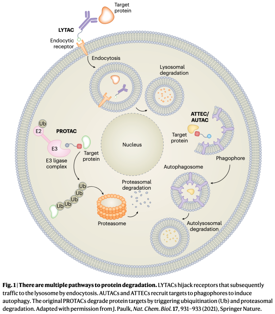
The new generation of degraders avoids the ubiquitin–proteasome system entirely and instead harnesses cellular pathways for delivering cargo to the lysosome for degradation. LYTACs, or lysosome-targeting chimeras, and ATACs, or ASGPR (asialoglycoprotein receptor)-targeting chimeras, link an extracellular domain of a target molecule to a cell-surface receptor that shuttles it, by endocytosis, to lysosomes. Genentech’s PROTABs, or proteolysis-targeting antibodies, and EpiBiologics’ AbTACs, or antibody-based PROTACs, hijack cell membrane ubiquitin ligases that also direct targets to lysosomes, and EpiBiologics’ KineTACs degrade extracellular and membrane proteins by tethering them to cytokine receptors for cell internalization. Several other degraders exploit natural autophagy pathways, in which specialized vacuoles engulf protein aggregates and complexes, or damaged organelles, for lysosomal delivery.

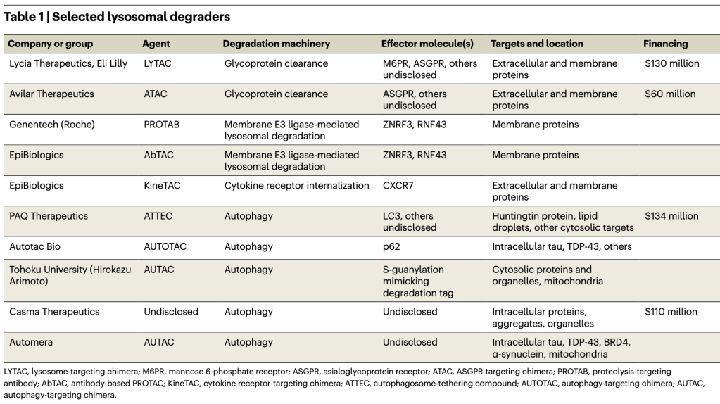
The field of lysosomal degraders is very young but is quickly growing. Besides LYTACs and the other extracellular protein degraders, at least five different autophagy-based degraders are in development (Table 1). At last count, five start-up companies have emerged from stealth mode, with over $434 million dollars invested. But while PROTACs have validated the degrader concept, lysosomal degraders are still untested in patients, and they face challenges and uncertainties as they advance to the clinic. These include oral delivery of bulky degrader molecules, the risk of off-target degradation, the potential to overload lysosomes, and the possibility that hijacking these pathways will disrupt cellular proteostasis.

Garbage in

LYTACs are the most visible new degrader modality. They can theoretically degrade most extracellular and membrane proteins, which together make up about 40% of the proteome. The first LYTACs originated in the Stanford lab of glycobiologist Carolyn Bertozzi, where Stanford University chemical biologist Steven Banik, then a postdoc, was looking for ways to eliminate proteins that crosslink the glycocalyx (the dense sugar-rich layer surrounding cells), which helps drive cancers. The Bertozzi lab had developed artificial glycoprotein polymers, and Banik planned to attach them to glycan-binding disease-related proteins and remove them, by shedding, from the cell surface. But eventually the duo hit on the idea of harnessing the cell’s natural machinery with PROTAC-like bifunctional molecules that operate in the extracellular space and, in combination with membrane-bound transporter molecules, grab extracellular proteins and shuttle them to the lysosome, where they are enzymatically digested (Fig. 1).

The initial LYTAC paper from the Bertozzi lab reported degradation of several therapeutically important proteins — the extracellular protein ApoE4 and membrane proteins EGFR, CD71 and PD-L1 — by linking them to the mannose 6-phosphate receptor, which internalizes and carries glycoproteins to the lysosome. Degrading membrane proteins was a feat because LYTACs must compete with these proteins’ normal internalization machinery. Banik, Bertozzi and colleagues later showed that degraders that used a second internalizing receptor, ASGPR, also worked, in this case in a tissue-specific manner. (Avilar Therapeutics’ ATACs are similar.)

In 2019 Bertozzi founded Lycia Therapeutics to develop and commercialize LYTACs. A $50 million A round followed, and in August of 2021 Lycia signed a research collaboration and licensing agreement with Eli Lilly. The companies have yet to disclose any targets or disease indications, except for Lilly’s focus on immunology and pain. “We’re looking for areas of biology and targets where antagonism has failed,” says Lycia CSO Steve Staben: “things like difficult-to-drug protein aggregates, immune complexes, some of these recalcitrant and ligand-independent membrane targets.” Autoantibodies are also attractive. Other possible targets include membrane proteins with multiple functions and interacting partners, making it necessary to completely remove the protein. Many receptor tyrosine kinases fall into this category, including cancer drivers EGFR, HER2 and FGFR. “A single degrader approach for one of those may be more efficacious than simple kinase domain inhibition or blocking natural ligand binding,” says Staben.

LYTACs enjoy more design flexibility than PROTACs because they don’t have to be cell permeant. Small molecules, peptides, monovalent antibodies, bispecific antibodies — almost anything works if it can bring the target to the receptor. “It’s important for us to have multiple modalities to target these extracellular proteins,” says Lycia president and CEO Aetna Wun Trombley. “You can imagine a fully small-molecule LYTAC that has quite a different pharmacological profile than a fully biologic LYTAC. And so, depending on the situation, it may be more beneficial to apply one of those modalities versus another.”

LYTACs are also relatively straightforward to design. In contrast, PROTACs must bind both targets in a ternary complex that enables the ligase to transfer ubiquitins to the target protein’s lysine residues. The lysines must be oriented properly for ubiquitin binding, and ubiquitination doesn’t always lead to proteasomal degradation. The rules for these interactions remain largely unknown, so drug discovery is empirical and expensive. LYTACs are more forgiving. “You just need to induce proximity transiently to the internalizing receptor,” says Staben. Once achieved, “there’s no need to maintain that ternary complex formation necessarily, there’s no absolute requirement for the suitable presentation of lysines.”

But unlike PROTACs, first-generation LYTACs do not act catalytically, so they may not be able to clear highly abundant proteins. Lycia isn’t ruling out catalysis in the future. “It’s more an engineering feat at this point,” says Banik. “We need to find something that’s going to let go of cargo in the lysosome while it itself can be recycled back to the cell surface.”

LYTAC toxicity, at this early stage, is unknown, but the general approach seems safe because it employs the cell’s normal endocytic machinery. However, the endosomal–lysosomal system is damaged in many diseases and chronic conditions, including neurodegenerative diseases, and the LYTAC cargo burden could make things worse. For example, impaired cargo degradation in lysosomes can lead to lysosomal membrane permeabilization and cathepsin release into the cytosol, inducing either apoptotic or necrotic cell death. “How a cell turns over its proteins is something that I think disease mechanisms tend to alter,” acknowledges Banik. However, “we have at least done initial profiling work to show that LYTACs themselves don’t really cause lysosomal damage in any way.”

Still, Lycia doesn’t want to overload sick lysosomes. “The different disease indications we’re looking at are not known to be associated with dysregulated lysosomal function,” Staben says. And selecting internalizing receptors with specific tissue expression should avoid any problems if issues do arise. “So it may be possible even in some of these patient populations that have lysosomal function changes, that we can avoid hypothetical on-target mechanism-driven toxicity.”

Avilar CEO Daniel Grau says the company hasn’t seen any safety issues in its preclinical studies, including testing in non-human primates. And, like Lycia, Avilar is staying away from diseases with known lysosomal damage or capacity issues. But degraders, by monopolizing the ASGPR (or any internalizing receptor), could theoretically block normal degradation and cause side effects. Grau cites a knockout study showing no reduction in plasma glycoprotein clearance in mice, suggesting that redundant systems are compensating. “There are a lot of backup pathways to degrade proteins,” says Avilar CSO Effie Tozzo. “Also … we’re definitely not saturating the receptor with our ATACs in our pipeline programs.” So degraders shouldn’t affect normal protein clearance. But, until safety is proven in the clinic, such interference remains a concern.

Both Lycia and Avilar are exploring multiple internalizing receptors, but Avilar has staked out a strong chemistry position with ASGPR. “They’ve disclosed quite a bit in terms of optimizing ligands for that receptor,” says Casma Therapeutics CSO Leon Murphy. “It’s impressive.” Unlike the original Banik–Bertozzi ligands, arrayed as bulky chains on their polysaccharide backbones, Avilar’s bypass the need for multivalency to activate ASGPR. “We can dispense with the requirement of having a multimodal presentation,” says Grau. Smaller size enables oral delivery. (Lycia, says Staben, also now has monovalent receptor ligands.) One limitation: while ASGPR degraders can target any extracellular protein, they can only degrade membrane protein targets expressed on liver hepatocytes, where ASGPR is expressed.

But the mannose 6-phosphate receptor and ASGPR are just two effector molecules. The total number of internalizing receptors and other trafficking proteins may be vast. “[There] could end up being hundreds of proteins that one might be able to think about using as a way to get to lysosomes quickly,” says Banik, whose Stanford lab is working to identify potential protein shuttles.

Another outside-in approach for degrading membrane proteins recently emerged from Wells’s lab: AbTACs. AbTACS are bispecific antibodies that place a transmembrane E3 ligase next to a membrane protein of interest, sending it to the lysosome for degradation. (Some E3 ligases sort proteins for degradation in the lysosome instead of the proteasome.) Genentech’s PROTABs used the same membrane E3 ligases to efficiently degrade several cancer-driving membrane proteins in cells. The Genentech team also identified 38 different membrane E3 ligases, many of them cell type specific, that future degraders might use to shuttle membrane targets to lysosomes.

But AbTACs and PROTABs may not be able to degrade extracellular (secreted) proteins, and, like PROTACs, they have tricky ubiquitination requirements, so proximity alone may not induce degradation. Wells’s KineTACs, or cytokine-targeted chimeras, don’t have those limitations, degrading the extracellular protein VEGF (vascular endothelial growth factor) in addition to membrane proteins. All these degraders expand on LYTACs “by both providing additional degradation platforms and tissue selectivity,” Wells writes in an e-mail. Both AbTACs and KineTACs were licensed to EpiBiologics in October.

Can autophagy deliver?

For intracellular proteins, PROTACs and molecular glues — small-molecule E3 ligase binders that alter that enzyme’s surface to recruit a target protein for degradation — remain the dominant degrader modalities. But their inability to degrade aggregates, non-protein targets and organelles has inspired a set of degraders that instead hijack the autophagy system, which efficiently clears such debris from the cell.

Autophagy, which means “self-eating,” starts when the phagophore, a cup-shaped vesicle, scoops up cell contents. The phagophore closes around its cargo to form the double-membrane autophagosome vesicle, which then fuses with the lysosome and deposits its contents there for digestion. “These are normal pathways that we can just enhance further … with a small-molecule degrader,” says Casma’s Murphy.

Companies see potential for autophagy-based degraders in neurodegenerative diseases, particularly those characterized by mutant protein inclusions (Huntington’s), by intracellular tau aggregates (Alzheimer’s), or by α-synuclein fibrils and damaged mitochondria (Parkinson’s). Targeting mitochondria with a degrader in Parkinson’s or any condition is perilous, but conceivable. “Having a disease-specific marker on a mitochondrion [would] likely allow for a favorable therapeutic window,” says Murphy, who says several groups are looking for such markers. Non-neuronal protein aggregation disorders like amyloidosis are also in the picture. Lipid droplets could be targeted in metabolic disease. Even soluble proteins, which are not typically cleared by autophagy, are potential targets, says Murphy. “The majority of proteins in the cell, at some stage of their life, are associated with complexes,” he says. “The target space is broader than what you might think.”

The autophagy degrader space is so new that the effector molecules, analogous to the E3 ligases that PROTACs employ, are mostly undisclosed by the companies testing them. PAQ Therapeutics, for example, is developing ATTECs, or autophagosome-tethering compounds, using undisclosed autophagy proteins. Boxun Lu, a neuroscientist at Fudan University in Shanghai, found the first ATTECs when he conducted a screen for small-molecule binders to mutant huntingtin protein, which accumulates in the brain in Huntington’s disease, and counter-screened against LC3, a critical protein that recruits cargo to autophagosomes for transport to lysosomes. This screen improbably produced two molecular glues that bound to both. Lu’s group went on to show that these compounds and their more target-specific analogs could reduce mutant huntingtin and reverse disease in cells and in mouse disease models. Lu originally set out to find heterobifunctional molecules, not molecular glues, so “it is an extremely lucky event they were able to identify these molecules,” says Nan Ji, CEO of PAQ Therapeutics, which has licensed the technology.

These compounds are now in PAQ’s portfolio, but finding more single-warhead molecular glues for any given target usually involves a massive screening effort. So the company is developing ATTECs with dual linkers, one to an autophagy effector protein and the other a target of interest. These are simple to construct. But unlike the similarly two-headed PROTACs, which can employ known and validated binders, autophagy targets are mostly unexplored territory, and there are few if any known small-molecule binders to autophagy-related proteins.

Nan Ji says that PAQ has begun to break that barrier. Besides finding target binders, “we have also been able to identify very potent small-molecule binders to an autophagy protein,” he says. “I can’t really specify which one yet.”

Other companies are also keeping their autophagy proteins of choice under wraps. There are many possibilities, but also pitfalls, because autophagy is more complex than the ubiquitin–proteasome system, with a lot more players. In budding yeast there are at least 41 autophagy-related (Atg) proteins, which are highly conserved in mammals. A single degrader molecule that binds only one must recruit the entire autophagy machinery. Also, several different mechanisms of selective autophagy exist, suggesting that a single degrader platform may not be possible.

Murphy acknowledges the challenge. “It’s not a classical signaling pathway or cascade — it’s just not,” he says. “There are multiple components that sort of feed in at different levels; there’s a lot of cooperativity and parallel requirements.” Success, he says, will come from acquiring knowledge of specific autophagy factors sufficient to assemble the machinery for degradation. “I do believe it is going to be possible, once in possession of that knowledge, to deploy that approach across multiple therapeutic targets,” Murphy says. A universal autophagy degrader platform, he says, is possible. “This concept of self-sufficiency really holds the key.”

One autophagy degrader approach is the AUTAC, or autophagy-targeting chimera. Fifteen years ago, a group that included Hirokazu Arimoto, an organic chemist now at Tohoku University in Japan, discovered S-guanylation, a post-translational modification of proteins. Arimoto later observed that bacterial pathogens accumulate S-guanylated proteins around them and are cleared by autophagy, suggesting a mechanistic connection. To make the first AUTAC degraders, he linked an S-guanylation tag to a protein of interest, driving its degradation by attaching ubiquitin chains joined at their lysine 63 residues, a pattern that typically triggers autophagy. AUTACs degraded not only cytosolic proteins but also dysfunctional mitochondria, improving cellular energy production.

What’s missing from the AUTAC story is an explanation for how the S-guanylation post-translational modification triggers autophagy. “There’s a gap there,” says Murphy. “It’s unclear exactly how that molecule interfaces with the autophagy machinery.” It’s not known whether AUTACs have been licensed for commercial development; Arimoto did not respond to email messages from Nature Biotechnology.

Casma, like PAQ, has not revealed which autophagy protein or proteins it plans to use to effect target degradation. Autotac Bio, which is developing AUTOTACs, or autophagy-targeting chimeras, has disclosed one: p62, which forms a bridge between a protein that’s marked for degradation and the autophagy machinery. p62 and other autophagy receptors are not classical drug targets. “Finding appropriately tractable small molecule binding sites is going to be essential to success,” says Murphy. Using LC3 could be problematic because recent work has revealed non-autophagy functions for this family of proteins, so recruiting them to degrade any given target could impair some normal biology — for example, the transcription of genes for antimicrobial defense.

A risk exists that autophagy-based degraders could interfere with proteostasis by appropriating the autophagy machinery. “In a chronic disease setting, that’s something to really pay attention to,” says Murphy. “Hopefully it’s something we can modulate, because we’re using chemistry and pharmacology, and we can adjust dosing schedules appropriately to minimize that risk.”

Safety aside, will autophagy degrader drug discovery run into the same unpredictability problem as PROTACs? “There are too many unknowns to say we will definitely have an easier path here,” says Murphy. Nan Ji agrees. On the one hand, he says, even though an autophagy degrader, just like a PROTAC, must form a target–effector ternary complex, “there would be no need necessarily for ubiquitination to occur. So in theory, that this would save one requirement for degradation to happen.” On the other hand, autophagy degraders may sacrifice some specificity in the process. With PROTACs, “because of the requirement for ubiquitination, the lysine positioning, you could get additional selectivity — degradation selectivity,” Ji says. With ATACS, “that may not be the case.” Less built-in selectivity could mean more optimization steps for autophagy degraders, or lower potency. PAQ is still working out the discovery process.

Other TACs and TABs will emerge. “There are many paths to the lysosome,” says Banik. PROTACs are almost certain to find a niche in medical practice — molecular glues like the multiple myeloma drug Revlamid (lenalidomide) already have. But whether targeted degradation will become a major drug modality at the level of small-molecule inhibitors and antibodies is much less clear. Even less certain is the ultimate fate of LYTACs, ATACs, AbTACs, KineTACs, PROTABs, AUTACs, ATTECs and AUTOTACs. They’ve barely gotten started, and their biology is mostly unexplored. Companies, just out of stealth mode, are only now beginning to disclose their clinical candidates and preclinical results. The next year will be telling.

Ref:

https://pubmed.ncbi.nlm.nih.gov/29626215/

First demonstration of miRNA-dependent mRNA decay

MicroRNAs (miRNAs) are important regulators of gene expression. Their function and roles were first discovered in the development of the worm Caenorhabditis elegans, and later shown to occur in all multicellular organisms. miRNAs function by guiding effector proteins through the recognition of complementary miRNA sequences in target mRNAs. In most animals, miRNAs form imperfect hybrids with sequences in the 3′ untranslated regions (3′UTRs) of mRNAs; the ‘seed’ region of the miRNA ensures targeting specificity. Nowadays, we know that this interaction leads to the recruitment of a protein complex that represses translation and causes deadenylation and degradation of target mRNAs.

By contrast, the original model that was proposed to explain the mechanisms underlying miRNA functions postulated that the mRNA remains stable after miRNA binding, and that gene repression occurs only at the level of translation. This conclusion was based on initial findings that miRNAs regulated the levels of target proteins, but not of target mRNAs. This model dominated the field until the publication of a key study by Amy Pasquinelli’s lab in 2005.

Pasquinelli and her team showed that two miRNAs in C. elegans — let-7 and lin-4 — trigger degradation of their imperfectly complementary mRNA targets (lin-41, lin-14 and lin-28). This conclusion was widely accepted thanks to the use of the same experimental system as the one used to establish the initial model and owing to the technical rigour of the work. The authors used northern blotting and reverse transcriptase quantitative PCR (RT–qPCR) to compare levels of miRNA targets across developmental stages, during which miRNA expression changes, and also between miRNA-mutant and wild-type worms. They ruled out transcriptional gene regulation as a mechanism of miRNA function using chromatin immunoprecipitation experiments. Finally, the authors confirmed mRNA degradation by carrying out lacZ reporter experiments with the wild-type 3′UTR of lin-41 or with a mutant 3′UTR, in which the let-7-binding sites were deleted.

“This work… triggered further mechanistic studies of miRNAs”

This work was followed up by numerous studies showing the degradation of miRNA targets in other organisms and triggered further mechanistic studies of miRNAs. Research in subsequent years provided an understanding of how miRNAs cause mRNA degradation and translation repression and how these two mechanisms contribute to overall miRNA-mediated gene repression.

References

Original article

  • Bagga, S. et al. Regulation by let-7 and lin-4 miRNAs results in target mRNA degradation. Cell 122, 553–563 (2005)

Probing RNA-Binding Proteins in Cancer

RNA-binding proteins (RBP) are an emerging class of potential cancer therapeutic targets, albeit with myriad complexities that have yet to be untangled. Recent research has implicated RBPs in MYC-driven cancers and in acute myeloid leukemia (AML)—two examples in a lengthening list.

“The early genomics era was mainly focused on DNA-level molecular mechanisms—transcription factors and epigenetic modifiers were seen as the drivers of gene expression control,” remarks Hani Goodarzi, PhD, of the University of California, San Francisco. “But some RNA biologists, including Robert Darnell’s group at The Rockefeller University [in New York, NY], were already looking ahead to how RBPs might also modulate gene expression.” Notably, Darnell developed cross-linking immunoprecipitation (CLIP) to pinpoint to which RNA type, and where, a given protein binds.

“Once we realized that transcriptional regulation is only one part of the equation, more researchers began paying attention to what happens after, in the post-transcription space,” Goodarzi adds.

Interest in RBPs picked up, and lately “the floodgates have opened,” notes Gene Yeo, PhD, of the University of California, San Diego, in terms of tools at RNA biologists’ disposal. These include mass spectrometry–based quantitative proteomics and STAMP, a technology his group developed to study RBP–RNA interactions in single cells (Nature Methods 2021;18:507-19). As well, CRISPR–Cas9 screening “has become very important for looking at synthetic lethality and function to better identify RBPs as novel drug targets.”

Occasionally, toolbox components are tweaked and refined; for instance, Yeo’s team came up with “enhanced CLIP”—dubbed eCLIP—which is less technically demanding, with improved specificity and success rates. eCLIP “enabled us to generate large-scale interaction maps of RBPs and their targets,” he says. “It turns out that there are considerably more of these proteins than previously thought; some 10% to 20% of the human genome encodes RBPs” (Nature 2020;583:711-9).

Another novel technique for mapping RBP networks, called HyperTRIBE, was adapted from studying nerve cells in fruit flies by Michael Kharas, PhD, of Memorial Sloan Kettering Cancer Center, also in New York, NY (Nat Commun 2020;11:2026). For Kharas, “the most powerful aspect of RBPs is their ability to really change the cell state, because they influence protein production, dictating how much or how little is made from a given transcript.” Cancer can interfere with this process, he adds, “tipping a delicate balance and thereby altering key decision points for the cell.”

“RBPs are complex molecules whose activities reverberate throughout the cell’s gene expression network,” Goodarzi concurs. Whereas transcription factors such as p53 have long received the spotlight as key regulators that tumor cells frequently hijack for their own purposes, “we’re learning that RBPs are master regulators, too, and also co-opted” in cancer. As well, “in sequencing more cancer genomes, we’re starting to uncover a ton of mutations in RBPs,” he notes, “which has really put them on the map for cancer biologists.”

Toward therapeutics

A recent study from Yeo’s group sheds new light on cancers addicted to MYC, which has long vexed researchers as a therapeutic target. However, probing the post-transcriptional milieu of MYC-driven tumor types may yield workaround strategies down the road (Mol Cell 2021;81:3048–64).

Yeo reported that YTHDF2, an RBP, is a vulnerability in triple-negative breast cancer (TNBC) with hyperactivated MYC. YTHDF2 typically keeps a lid on the number of mRNA transcripts that are translated, earmarking many for degradation to maintain cellular homeostasis. With MYC addiction, transcription and translation levels are aberrantly high, so YTHDF2 “becomes more important than ever” for balance, he explains. “When we inhibited it, that provoked a lot of cellular stress from accumulated unfolded proteins, which then triggered apoptosis” in TNBC cells and tumor xenografts.

“Our findings show how cancer cells exploit the function of specific RBPs, to evade stress-induced death,” Yeo adds. “To us, YTHDF2 is a plausible therapeutic candidate, but of course there are others out there.”

“Others” may include RBMX and RBMXL1, which Kharas and his team have been studying. After initially identifying the RBP Musashi-2 as an important regulator in AML, they began scoping out Musashi-2′s network, landing on RBMX and RBMXL1. Both are overexpressed in AML and necessary for tumor cell survival (Nat Cancer 2021;2:741-57).

“We found that these two RBPs directly promote the transcription of their target, CBX5, itself a regulator of chromatin accessibility in AML cells,” Kharas explains. “Knocking them out reduced CBX5′s mRNA and protein abundance, changing how chromatin is compacted, which stunted cell growth and delayed leukemia development.”

The number of identified RBPs is estimated at 1,500, so deciding which ones to pursue therapeutically “will come down to prevalence,” Goodarzi says. “If an RBP’s mode of regulation is pretty extensive, impacting a broad set of cellular and cancer states, that opens the door for it to be prioritized.”

For instance, SF3b1—a key RNA splicing component—is frequently mutated in patients with myelodysplastic syndromes, which can morph into leukemia. H3B-8800 (H3 Biomedicine), a small molecule that modulates SF3b1′s activity, is one drug being evaluated in the clinic. However, in preliminary data from a phase I trial of 15 patients there were no objective responses (Leukemia 2021 Jun 25 [Epub ahead of print]).

A challenge is that RBPs “have different functions that are wholly context-dependent; the same protein that’s a splicing factor in the nucleus can be a stability factor in the cytoplasm,” Goodarzi says.

Yeo agrees: “You’d need to discern what, exactly, to target—is it an RBP’s RNA recognition function, its ability to recruit other proteins as part of a complex, or something else entirely?” Compounding the complexity, many RBPs have intrinsically disordered regions, “which are structurally unstable and can form aggregates. We don’t yet know if this aspect would make them easier or harder to drug.”

Strategies being explored include not only small-molecule inhibitors, but antisense oligonucleotides that modulate RBPs at their own transcript level. As well, decoy RNAs conjugated to proteolysis-targeted chimeras (PROTAC) could selectively trap RBPs, routing them toward degradation. This RNA–PROTAC concept has shown utility in vitro, targeting LIN28 and RBFOX1 in cancer cell lines.

“The more we learn about RBPs, and with better technology, the fancier we can get in thinking about how to drug them,” Kharas says. “This is just the beginning.”

“I’d say RNA is having a renaissance moment,” Goodarzi adds. “We understand very little about post-transcriptional control. It’s this vast landscape, and we’ve barely scratched the surface. But the tools keep improving, and excitement in the field is driving participation, so now is a pretty great time to be an RNA biologist.” –Alissa Poh

Edge/Chrome浏览器的扩展 推荐:2022版

1. View Image: 右键执行以图搜图功能
2. Scholarscope : 显示pubmed影响因子(需付费)
3. IDM Integration Module
4. Quillbot
5. SimpRead – Reader View
6. Tampermonkey
7. uBlock Origin
8. Blink: Organize your work in progress
9. Clip to DEVONthink: clip selected web content to your DEVONthink database.
10. Fiteprint – Current site history: Quick access to current site history.
11. Infy Scroll: Add customized infinite scrolling to websites and auto-load the next page. Supports the AutoPagerize Database
12. Save image as Type: Save image as PNG, JPG, or WebP by context menu on the image.
13. Science Research Assistant: 右键搜索pubmed或者scholar
14. Shipped and sold by Amazon: Show only products that are shipped and sold by Amazon.com
15.Shortcuts for Google:
16. keepa:亚马逊历史价格追踪

病毒载体的基本元件及其优化

一. 启动子

启动子是可以启动目的基因转录的DNA序列,该序列可以被RNA聚合酶所识别,并开始转录合成RNA。启动子可以和调控基因转录的转录因子产生相互作用,控制基因转录的起始时间和转录的强度,它就像 “开关”一样,决定基因的活跃程度,继而控制细胞开始生产哪一种蛋白质。目前绝大多数的基因治疗产品,都的是病毒启动子。这主要是还是从提高蛋白表达效率的角度出发,希望尽可能地利用少量的病毒去表达尽可能多的目标蛋白。而病毒的启动子往往是强的组成型启动子,他们可以在宿主细胞内,招募宿主的转录因子,达到比宿主启动子高得多的转录水平。并且在长期的进化中,病毒的基因组结构已经变得十分紧凑,长度较短,非常适合应用于基因治疗载体。

目前最常见的病毒启动子是人巨细胞病毒的早期启动子(CMV-IE promoter/ CMV promoter),在CMV中,这个启动子负责IE1 和 IE2 基因前体 RNA 的转录起始。最常用的是一个600-800bp的CMV enhancer/promoter/UTR的融合版本,它能够在许多的组织中达到一个高强度的转录水平。但也有报道称,CMV 启动子驱动的蛋白表达水平会随着培养时间的延长而降低,宿主细胞会提高DNA甲基化的水平,从而将CMV启动子的转录沉默掉。

还有一些人工组合的启动子(有些也融合了也包含了部分intron序列在里面)也利用了CMV的部分序列,例如CAG(C,the Cytomegalovirus early enhancer element;A,chicken beta-Actin promoter; G, splice acceptor of the rabbit beta-Globin gene)、CBh启动子等等。CAG启动子的全长大约有1.8kb,’’结构较为复杂,具有非常强的转录活性和广泛的宿主范围。但由于序列过长,因此在应用于AAV时,具有明显的局限性。于是一些实验室在这个基础上开发了类似的混合启动子,在缩小了启动子尺寸的同时,大体保证了它的转录活性。例如简化的CAG/CBA启动子,简化后的序列长度仅有584bp。除了这些常见的病毒启动子之外,还有很多管家基因的启动子,也被用于基因治疗当中。比较普遍的包括1.2kb 的EF1-α 启动子、500bp 的PGK 启动子以及UBC启动子等等,但是其转录能力,就要弱于CMV、CAG等高强度的启动子们。

当大家只想在某些特定的器官或者特定类型的细胞内表达目的基因时,就需要使用组织特异性的启动子,或者给特定启动子加上一些组织特异性的调控原件。这样即使在载体没有特别好的靶向性的情况下,也可以限制目的基因在非靶标组织的表达泄露。例如胶质纤维酸性蛋白 (GFAP, 2.2 kbs) 启动子以及截短版本的gfaABC(1)D都被用来在星形胶质细胞中进行特异性的基因表达。此外还有在肌肉中常用的MCK,心肌中常用的cTNT,肝脏中常用的hAAT、ApoE等。但这些启动子也有很难克服的局限性:表达量低和尺寸较大。这是组织特异性表达需要给顺式作用元件提供大量的序列空间,因此其尺寸就必然要大于组成型启动子;另外组织特异性启动子需要被特定组织细胞内有限的反式作用因子所调控,即其所能被上调的峰值也是较低的。目前也有很多实验室通过生物信息学的方法来分析启动子及其调控原件的序列,精简或者串联多个不同来源的调控原件,以增强组特特异性表达的强度,也获得了一些喜人的突破。

在真核细胞中,有三种不同的 RNA 聚合酶,即 RNA 聚合酶 I、II 和 III。RNA 聚合酶 I 合成了大多数的 rRNA, RNA 聚合酶 II 转录所有 mRNA 和许多非编码 RNA,RNA 聚合酶III 则转录了其他的小的非编码(5S rRNA, snRNAs, snoRNAs, SINEs, 7SL RNA, Y RNA, 以及 U6 spilceosomal RNA等等)。如上所述,用于目的基因表达的启动子都是结合RNA 聚合酶 II,但如果基于想在病患体内表达功能性RNA例如shRNA、miRNA、sgRNA等等,这个时候就需要用到结合RNA 聚合酶 III的 III型启动子,目前最常用的有小鼠的U6启动子和H1启动子。而在让目的mRNA转录终止时,通常使用 T-stretch 作为终止信号,但终止效率和实际终止位点也并不是一致的。其中T4 信号是最小的终止信号,但完整的转录终结只有在 T-stretch ≥6 时才能达到。如果没有达到完整的转录终结,可能会产生低水平的 3′ 延伸 RNA,进而会干扰下游基因的表达。因此,在设计小 RNA 表达盒中至少应使用可以实现完全 Pol III 终止的T6/7 信号。

二. 转录后调控原件

在表达外源基因时,将一些转录后调控原件插入到3′ 非翻译区,可以增加mRNA在细胞内的积累水平并提高翻译效率,这样也可以在一定程度上增加外源基因的表达量。目前比较常用的是乙型肝炎病毒的转录后调控元件 (HPRE) 和土拨鼠肝炎病毒的转录后调控元件 (WPRE)。它们都是嗜肝DNA病毒的顺式作用 RNA 元件,可以通过促进 mRNA 从细胞核输出到细胞质来增加mRNA在细胞质 内的积累,增强3’端加工效率和mRNA稳定性。通常,未经剪接的 mRNA 会以较低的效率运输到细胞质中,然而一些细胞因子可能与 PRE 元件相互作用并介导其转录后的转运(RNA在核仁中转录后,会经过快速的加工过程,去除翻译的间隔区,产生成熟的mRNA,这些mRNA可以与核糖体结合并在细胞质中大量积累 。虽然前体RNA 的半衰期非常短~mins,但成熟的mRNA在细胞质中的半衰期则会较长~days。因此,帮助mRNA从核仁到细胞质的运输,在一定程度上提高了细胞内mRNA的积累水平)。然而,尽管 HPRE 、WPRE 对基因表达有益,但它们的序列相当长(600 bp),因此只能用于其他表达元件尺寸很小的表达框中。

三. polyA加尾信号

终止子位于基因序列的3’末端,通常直接出现在3′ 调节元件之后,它可以将新合成的mRNA 从转录复合物中释放出来。虽然启动子强度是决定基因表达水平的主要因素,但终止子在 RNA 加工中也发挥着重要作用,它会直接改变RNA 的半衰期,并最终导致基因表达水平的改变。目前常用的终止子包括SV40、hGH、BGH 和rbGlob,他们在序列上都包括促进多聚腺苷酸化和终止的motif AAUAAA。mRNA的转录终止和多聚腺苷酸化是一个协同过程,mRNA会在polyA加尾信号AAUAAA与下游的GU-Rich区之间发生切割,从而产生一个游离的 3′ 末端并用于在此基础之上进行polyA加尾。

四. 多顺反子载体—-一根藤上七个瓜

人类大多数的疾病并不是简单的单基因病,在治疗时,科研人员往往需要同时给与患者多种蛋白分子进行共同作用,因此基因治疗还要面对需要同时递送和表达几个目的蛋白的挑战。将多个基因克隆到一个载体中,从而进行多个基因的联合表达会在一定程度上提高基因治疗的有效性。目前常用的策略是在表达框中加入内部核糖体结合位点IRES或者加入有自切割功能的2A Peptides。

大多数情况下,真核生物的翻译起始需要mRNA的5’帽子与核糖体的小 (40S) 亚基以及许多翻译起始因子 (eIF) 相结合。而IRES 序列可以控制不依赖于5’帽子的蛋白质合成。IRES 序列大多是在病毒中发现的 ,大小多长于500 bp,目前最常用的是EMCV 和MSCV的IRES。

2A 肽是源自病毒的短肽 (~18-25 aa)。它们具有自我切割的功能,可以从同一个转录本中表达出多种蛋白质。但实际上,2A 肽的自我切割,并不是完整的把2A多肽切割下来,甚至它就不是真正的对翻译好的肽链进行切割。以源自口蹄疫病毒的2A 肽F2A为例-(GSG) VKQTLNFDLLKLAGDVESNPG P,F2A的功能是使核糖体跳过2A 肽 C 末端甘氨酸G和脯氨酸P之间肽键的合成,从而导致 2A 肽末端和下游肽段分离成两条多肽。因此,上游蛋白的 C 末端会添加额外的 2A 残基即(GSG) VKQTLNFDLLKLAGDVESNPG,而下游蛋白的 N 末端会添加额外的一个脯氨酸P。目前常用的2A肽有四种,P2A、T2A、E2A 和 F2A,它们来自于四种不同的病毒。

IRES 的主要缺点是两段蛋白的表达水平不一致,与多顺反子中的上游 ORF 相比,IRES 下游ORF 的表达水平会低很多(通常为上游表达水平的 10-20%)。IRES 元件也可能由于其尺寸(>500 bp)的问题,增加病毒包装的难度。2A 肽的缺点则是两个ORF 上留下的2A肽残基可能会影响目的蛋白的活性。此外,2A 肽的自切割不是 100% 有效的,并且切割效率会受到上下游 ORF 序列的强烈影响。因此,来自多顺反子的大部分翻译产物可能是未能自切割的融合蛋白,这在很多的应用中会是一个重要问题。在四种常用的 2A 肽中,P2A 通常具有最高的切割效率。接下来是 T2A,然后是 E2A 和 F2A。F2A的裂解效率仅为50%左右。

五. 表达框内的其他元素—-内含子与UTRs等

内含子的存在,可以影响包括转录、多聚腺苷酸化、mRNA 输出、翻译效率和 mRNA 降解在内的基因表达的各个步骤。然而前体RNA的剪接其实是一个非常耗能的步骤,剪接体内含子的切除需要在复杂剪接体的帮助下才能完成剪接过程(剪接体甚至是细胞中最大的分子复合物之一)。因此在长期的进化中,内含子的存在对细胞是有巨大负荷的。内含子对生物真正的意义,还有待于人们去发掘。而在设计基因表达框时,引入一些特别的真核生物内含子可以增加目的基因的表达量,这些内含子在转录起始位点的下游发挥作用。另外mRNA 的非翻译区 (UTR) 内存在多个调控元件,这对于 mRNA 的稳定性和蛋白翻译的效率也是非常重要的。譬如说 β-珠蛋白的5′-和 3′-UTR 可以明显提高翻译效率, α-珠蛋白的 3′-UTR则可以稳定mRNA,非洲爪蟾 β-珠蛋白 5′- 和 3′- UTR 、TEV的5′-UTR和人热休克蛋白 70 的 5′-UTR等同样被发现可以提高mRNA 的翻译效率等等。另外,在有些研究中,与内源性 miRNA 互补的靶序列也被整合到了表达盒里,这样可以起到在特定miRNA高表达的组织中“de-target”或抑制转基因表达的作用。

* (GSG) 残基可以添加到肽的 5′ 末端以提高切割效率

设计一个合格的基因表达框是一份可以很简单但又可以很复杂的工作,对不同组件的挑选、组合与改造,将会决定外源基因在目标组织中的表达量与表达时间。而AAV较小的外源基因承载空间,则给研究人员的发挥限制在了一个较小的舞台。如何在螺蛳壳里做道场,还是要靠科研人员针对不同的外源基因、靶组织进行case-by-case的尝试,这样才能开发出优质的表达框。

此外,所有蛋白表达框DNA序列都不是孤立的,必须构建到质粒骨架上(如AAV载体),与质粒骨架组成一个完整的闭环质粒,这对于DNA长期稳定的保存及后续的扩增生产都是至关重要的。

Gene-therapy innovation: Unlocking the promise of viral vectors

The past year revealed both successes and setbacks for viral-vector gene therapies. The rapid development and large-scale rollout of multiple adenovirus-vector vaccines represented an unprecedented achievement that is poised to help mitigate the devastating impact of the COVID-19 pandemic. During the same period, multiple high-profile gene-therapy assets encountered challenges, with clinical trials paused because of safety concerns or failing to meet efficacy targets.

These successes and setbacks are emblematic of the current state of viral-vector gene therapy: a technology with considerable promise but with a set of challenges still ahead. As more and more gene therapies have reached the clinic, it has become clear that multiple technological challenges must still be overcome to unlock the full potential of viral-vector gene therapy.

Rising to meet these challenges, biotech and pharmaceutical companies are testing a multitude of technological advances and innovative strategies that address all aspects of viral-vector gene-therapy development. For companies prepared to keep abreast of the rapid pace of change, these innovations offer a path for ushering in the next generation of viral-vector gene therapies.

The state of viral-vector gene therapy

Viral-vector gene therapies use modified viruses as drug-delivery vehicles to introduce specific DNA sequences—encoding genes, regulatory RNAs (for example, small interfering RNAs [siRNAs]), or other therapeutic substrates—into cells. The technology has long drawn interest for its potential advantages over traditional modalities. Many types of therapeutic agents (for example, enzymes, antibodies, and siRNAs) can be encoded in DNA sequences that can be rapidly designed and synthesized once a target is identified.

Viruses serve as powerful delivery vehicles for these sequences because of their ability to enter cells efficiently and potentially gain access to hard-to-reach, highly specific cells. In combining these features, viral-vector gene therapies can be used to modify gene expression in a programmable way, offering the flexibility to potentially treat a wide spectrum of diseases—including rare monogenic diseases by gene replacement and broad-population diseases by controlling gene expression—and help disease prevention by immunization.

Nearly all gene therapies currently available use one of three vector types: adeno-associated-virus (AAV) vectors, adenovirus vectors, or lentivirus vectors (Exhibit 1). AAV and adenovirus vectors are typically used in gene therapies that are directly administered to patients by infusion or local administration (in vivo), with AAV being the most popular vector for areas outside of oncology and vaccines. Lentivirus vectors are typically used for ex vivo therapies, in which cells harvested from a patient are modified in the lab before retransplantation. This article primarily focuses on in vivo gene therapies; however, many of the challenges and advances discussed are applicable across both routes of administration.

Excitement around viral-vector gene therapies is evident. While only four in vivo viral-vector gene therapies are currently on the market, more than 100 gene-therapy assets are in clinical trials as of late 2020, with a far greater number in preclinical development.

Many of these assets have emerged from the steady stream of small- and midsize biotech companies and academic labs supported by continued, high levels of venture-capital funding. Large pharma companies have increasingly focused on the potential of viral vectors, with seven biotech-company acquisitions valued near or above $1 billion in the past two years alone (Exhibit 2).1 Adenoviruses are being proven as a vaccine platform, with approvals for Ebola vaccines and groundbreaking COVID-19 vaccines over the past year.

While the high list-price of some gene therapies was once seen as a near insurmountable challenge to commercialization, innovative reimbursement strategies have shown that successful launches are possible, with ZOLGENSMA (treating more than 600 infants with spinal muscular atrophy3 in its first ten months on the market) beating analyst expectations.4 Worldwide sales of viral-vector gene therapies are forecast to grow at a rate of more than 50 percent year-on-year for the next five years (excluding the potential impact of COVID-19 vaccines), affecting the lives of tens of thousands of patients.

However, while there is significant momentum, there have also been multiple recent setbacks.5 Many of these relate to challenges previously outlined by McKinsey in its perspective on the future of gene therapy (including efficacy, durability, and manufacturing). As these therapies have sought to expand beyond the ultrarare indications they originally targeted, three technological challenges have emerged as recurrent obstacles. For viral-vector gene therapies to reach their true transformative potential—much like monoclonal-antibody technology 20 years ago—this set of technological challenges must be overcome.

Challenges to realizing the potential of viral-vector gene therapies

The current generation of viral-vector gene therapies represents the culmination of decades of biological and clinical research. As more patients have received these therapies, it has become clear that three fundamental challenges will restrict the applicability of viral vectors: getting past the immune system, lowering the dose, and controlling transgene expression. Ongoing work to address these challenges is generating technological innovations that have the potential to leapfrog current therapies and unlock the potential of viral vectors.

1.Getting past the immune system

The success of any viral-vector gene therapy depends on its ability to get past multiple lines of defense deployed by the human immune system. Viral capsids, viral-vector DNA, and even the transgene products themselves may be recognized as foreign, providing multiple opportunities for the immune system to clear the gene therapy from the body.

Immunity against viral capsids can limit the efficacy of a gene therapy. Because most viral-vector gene therapies today use vectors derived from harmless viruses circulating in humans, many patients (up to 60 percent) may have preexisting immunity from past exposure.6 CanSinoBIO, for example, reported reduced efficacy of its COVID-19 vaccine in individuals with preexisting antibodies to the adenovirus-5 (Ad5) vector it chose for drug delivery.7
Although this effect depends on the vector serotype used, and the clinical impact is still unclear,8 many clinical-trial sponsors conservatively exclude patients from their studies if they have antibodies to the vector in question. This can come at the cost of making most patients ineligible for therapy. Acquired immunity to viral vectors poses additional challenges for viral-vector gene therapy in the long term. Patients treated with a gene therapy today may not be able to receive a second gene therapy in the future if the same viral vector is used in both contexts.

In addition, viral capsids and viral-vector DNA can actively provoke an immune response from the body. For viral-vector vaccines, this immunogenicity can be beneficial, as it reduces the need for adjuvants and increases efficacy. However, for other viral-vector gene therapies, immunogenicity can reduce efficacy, increasing the chance that the gene therapy is detected and eliminated by the immune system. Indeed, some have speculated that immunogenic vector DNA sequences are behind the limited durability of some recent gene therapies, leading to their abandonment.9 More concerningly, immunogenicity can lead to safety concerns during therapeutic use, as high levels of viral capsids can cause severe immune reactions at the time of injection.

Unraveling the immune system’s intertwined responses to viral-vector gene therapies remains difficult. Animal models do not recapitulate all relevant aspects of the human immune system (as immune systems behave quite differently among species). While human clinical trials offer a valuable source of insight, many gene-therapy trials are too small to confidently isolate the parameters associated with a drug’s success or failure.

2.Lowering the dose

Current viral-vector gene therapies require the administration of large numbers of viral particles to patients, particularly for therapies aimed at treating systemic diseases. For example, recent gene therapies for Duchenne muscular dystrophy (DMD) that aim to correct mutations in muscle cells throughout the body have delivered up to approximately 10^16 (ten-thousand trillion) viral particles in a single dose (for example, a dose of 3 × 10^14 vector genomes [vg] per kilogram [kg], assuming a 30-kg child),10 which is multiple times the number of cells in the human body.11 For systemic diseases, the need to individually target and repair many cells in the body partly explains why such large doses are administered. Another explanation is the limited cell-type specificity of current viral vectors: large numbers of viral particles must be delivered to ensure that an adequate number reach clinically relevant cells.

The large doses used in current gene therapies pose two challenges. First, large doses are difficult and expensive to manufacture. Today, a typical manufacturing run of an AAV-vector therapy using high-yield cell lines and large-capacity bioreactors might only produce approximately ten doses of a systemic gene therapy from a single batch at a cost of nearly $100,000 per dose (assuming approximately 1 × 10^17 vg per batch).12 Although these costs will gradually decrease as gene therapies begin to reach clinical and commercial scales, any technological advance that reduces the required dose would bring immediate benefit, as a tenfold reduction in dose might also bring about a tenfold reduction in costs.

Second, and even more critically, administering large doses of virus has been linked to adverse safety outcomes.13 Although investigations of four deaths in clinical trials of AAV-vector therapies in 2020 are ongoing, three deaths occurred in high-dose cohorts. Clinical-trial protocols have subsequently been revised to limit viral dosage, reflecting the tremendous importance of this issue.[14]

3.Controlling transgene expression

Once a viral vector successfully delivers its therapeutic gene to the cells in question, the efficacy of the gene therapy depends on the quality of transgene expression. Specifically, the transgene must be expressed at the appropriate level (neither too low nor too high), in the appropriate cells, and for the appropriate duration to mediate the desired clinical effect. For therapeutic uses (in contrast to use for vaccines), the transgene may need to be expressed permanently if the gene therapy is to serve as a one-time cure and represent an appealing alternative for patients over current standards of care requiring repeated dosing (which may not be possible because of the challenges previously laid out). Regulators have required multiple years of follow-up data showing that gene expression is maintained. Indeed, some drugs have been abandoned when expression waned after 12 months.

To maximize chances of success, early viral-vector gene therapies have opted to include regulatory elements (DNA sequences such as promoters and enhancers that control how genes are expressed) that have been selected to drive high levels of transgene expression in all cell types. However, this approach may have significant drawbacks, particularly as gene therapies move beyond gene replacement for monogenic rare diseases. Overexpression of the transgene or its expression in the wrong cells may contribute to inflammation and other toxicities (as was observed in recent studies of nonhuman primates).15 Moreover, current gene therapies, once administered, cannot be controlled or turned off by clinicians should the need ever arise.

Innovative solutions that address gene-therapy challenges from many angles

To tackle the challenges facing gene therapy, academic labs, start-ups, and established companies are generating myriad innovative solutions (Exhibit 3). Each focuses on a specific component of a gene-therapy product (for example, the viral capsid) or part of the development process (such as manufacturing). However, these innovations often address multiple core challenges, outlining multiple paths to realizing the promise of viral-vector gene therapy.

We have identified five key trends to watch.

1.Improved capsids
The viral capsid is a critical component of viral-vector gene therapy. It determines which cells are targeted, the efficiency of cell entry, and the probability that the gene therapy is detected and eliminated by the immune system. In addition, the capsid is largely responsible for the stability of the viral vector during the manufacturing process and can affect storage and distribution requirements.16

The capsids most widely used today, including those used in on-market products, are derived from naturally occurring viruses. They have suboptimal properties, including little cell-type specificity, moderate efficiency of cell entry, and relatively high levels of preexisting immunity in humans. To address the problem of preexisting immunity, many assets use capsids from viruses found in other species. For example, the AAV8 and AAVrh74 capsids used in multiple AAV-vector gene therapies are derived from AAV serotypes isolated from macaques, and some of the COVID-19 vaccines that have been developed have used adenovirus serotypes from chimpanzees and gorillas. While this approach may limit the challenges of preexisting immunity, it largely doesn’t address specificity or efficiency (particularly as these viruses have evolved to infect nonhuman species).

Increasingly, drug developers are turning to capsids that have been engineered in the lab and can be selected to overcome the challenges mentioned previously (Exhibit 4). These engineered capsids are identified through large-scale screening efforts in which millions of variant capsids are screened for the desired properties and iteratively refined. Capsid-engineering platforms—many of which have been spun out of academic labs to form companies—achieve these ends by leveraging advanced technologies, such as cryo-electron microscopy (cryo-EM) and artificial intelligence.

Improving capsid properties could bring multiple immediate benefits. For example, a twofold increase in a capsid’s cell-type specificity could enable a twofold decrease in the overall viral dose required, thereby improving safety and cost. It’s still too early to determine the true impact of capsid engineering, as most engineered capsids are still in preclinical development. However, companies’ early reports suggest that capsids with five- to tenfold improvements in multiple attributes may be entering the clinic soon.

2.Improved vectors
Like the capsid, the DNA sequence of the viral vector itself affects multiple aspects of a gene therapy’s performance, but engineering the vector can often be considerably easier, cheaper, and quicker. Accordingly, vector engineering is becoming a growing focus of gene-therapy R&D. Vector engineering is often easier with adenovirus and lentivirus vectors than with AAV vectors because of AAV’s inability to package large pieces of DNA. However, innovative vector elements are beginning to appear in AAV-vector designs as well.

Vector engineering broadly has two aims: reducing the immunogenicity of the viral vector and improving transgene expression. One strategy to achieve both aims is codon optimization, in which variations in the vector sequence are explored to eliminate immunogenic sequence motifs while optimizing the transgene for robust expression. Subtle changes in vector sequence achieved through codon optimization can have large effects, such as increasing expression levels and possibly extending the duration of expression for multiple years.[17]

Transgene expression can be further programmed by engineering regulatory elements into the vector sequence. Some regulatory elements turn on transgene expression only in certain cell types or tissues—ideally, the disease-causing cells—preventing potentially toxic expression in other contexts. Such cell-type- or tissue-specific regulatory elements (for example, promoters and enhancers) have become relatively common in viral-vector gene therapies. For an additional layer of control, some viral-vector gene therapies are also incorporating regulatory elements, such as microRNA-target sites, that reduce expression in specified cells—for example, in cells that promote an immune response.

Finally, a more distant and challenging goal is to engineer vectors that are inducible, where transgene expression can be controlled using an additional signal, such as an orally administered small-molecule drug. This could allow clinicians to turn on, turn off, or otherwise adjust a gene therapy after it is administered, delivering a personalized course of treatment.

3.New types of cargo

The cargo delivered by a viral-vector gene therapy is typically a working copy of a gene that is used to replace the patient’s disease-causing copy of that same gene. However, any therapeutic agent that can be encoded in DNA can theoretically be delivered by a viral vector. Researchers and drug developers are increasingly leveraging this flexibility to deliver other types of molecules with therapeutic value—alone or sometimes in combination—including regulatory RNAs (for example, short hairpin RNAs [shRNAs]), vectorized antibodies, and substrates for gene editing.

Gene editing is an intriguing potential solution for achieving long-lasting, physiologically appropriate gene expression. For patients with diseases caused by certain types of mutations, restoring the function and expression of the patient’s own copy of the gene through gene editing may be simpler (and more permanent) than attempting to engineer and deliver a replacement.

4.Improved manufacturing processes

Early gene-therapy-manufacturing processes originated in academic labs and were focused on small, research-scale batches. These processes were not optimized for moderate- or large-scale production or for the delivery of systemic therapy. As gene therapies start to expand outside the treatment of ultrarare diseases, one of the many challenges being addressed is the presence of empty capsids created during the manufacturing process. These empty capsids, which have no active cargo, can create the requirement for higher doses and, accordingly, stimulate stronger immune responses.

Two approaches are being developed to reduce the ratio of empty-to-full capsids in manufacturing: developing improved methods to separate the empty from full capsids based on specific properties (for example, charge and molecular weight) and engineering cell lines that package full capsids more efficiently. By reducing the empty-to-full ratio, these advances reduce manufacturing costs, reduce immune responses, and improve the safety of gene therapy. Indeed, regulators have used reducing the empty-to-full capsid ratio as part of the rationale for lifting clinical holds on gene-therapy products with previous safety issues.[18]

5.Improved pretreatment and conditioning regimens

Beyond engineering the capsid and vector, a separate approach for reducing the immune system’s detection of viral-vector gene therapies involves coadministering the therapy with an immunosuppressive agent. Multiple such conditioning regimens are currently being tested to reduce the impact of neutralizing antibodies on the efficacy of the treatment, both of preexisting antibodies and newly generated antibodies that could prevent future redosing. Nearly all current viral-vector gene therapies use steroids to help manage the potential immune response to the viral vector; however, the type, dosage, and timing of the steroid treatment varies widely.

Some clinical trials are experimenting with more targeted immune suppression, such as the use of rituximab to reduce the creation of memory B cells.19 An even greater assortment of approaches is being tested in animal models to directly reduce the presence of neutralizing antibodies. These include the use of enzymes cleaving to immunoglobulin G (IgG), plasmapheresis to remove the neutralizing antibodies specific to the gene therapy, and even CRISPR-based repression of neutralizing-antibody creation.20 These approaches could expand the pool of eligible patients to include those with preexisting immunity. Moreover, these approaches could enable a patient to receive multiple doses of the same therapy or of different therapies using the same vector backbone.

The road ahead

Viral-vector gene therapies find themselves at another inflection point. Early successes in the treatment of rare diseases and vaccines have proven the potential of this modality, while the challenges to gaining widespread adoption—the way that monoclonal antibodies have over the past 20 years—have only become clearer. Nevertheless, the wealth of innovative solutions being explored across academia, biotech, pharma, and contract development and manufacturing organizations demonstrate that viral-vector gene therapies are here to stay.

As described previously, different solutions are emerging to address each of the core challenges. The diversity of these approaches and the complexities of gene therapy mean that no single approach is likely to “win.” That situation will enable a rapid innovation cycle in which gene therapies are constantly being improved upon, which will offer new opportunities to leapfrog existing products. Even as AAV-vector-based delivery is becoming the leading technology, some prominent limitations combined with the rapid pace of innovation leave the door open for other delivery technologies to emerge.

Owners of viral-vector platforms will need to consistently look to the next set of innovations beyond their current platforms and assets. That could include investing directly to help overcome the broader challenges or buying or licensing critical technology to upgrade their platforms. Indeed, multiple new biotech companies have launched to solve one or more of the challenges outlined in this article as a service to developers of gene therapies. Staying abreast of these developments will require fastidious monitoring of scientific and technological progress on all fronts. However, since it is difficult at this early stage to place bets across all potential solutions and innovators, gene-therapy leaders will need to make their investments judiciously.

In the short to medium term—while technological challenges limit the scope of gene therapies to curative treatments for rare diseases—fast followers may find it difficult to be successful, even with improved technologies, as first entrants rapidly address prevalent populations. Gene-therapy leaders will therefore need to strike a careful balance by accelerating programs today while retaining the flexibility to adopt innovative technologies that unlock treatments for broader-population diseases and the full promise of viral-vector gene therapies in the long term.

https://www.mckinsey.com/industries/life-sciences/our-insights/gene-therapy-innovation-unlocking-the-promise-of-viral-vectors

Circular RNA Translation

Definition
A new RNA family has emerged, circular RNAs (circRNAs), generated by a process of backsplicing. CircRNAs have a strong impact on gene expression via their sponge function, and form a new mRNA family revealing the pivotal role of 5′ end-independent translation. CircRNAs are translated into proteins impacting various pathologies including cancer and neurodegenerative diseases, and are key players in aging. RNA circle translation also provides many perspectives for biotechnological and therapeutic applications.

Introduction
The potential of circular RNA to be translated has been studied since the 1970s. In 1979, an experiment was designed to determine the ability of circular mRNA to attach ribosomes [1]. A synthetic RNA was circularized with T4 RNA ligase and the binding of bacterial 70S ribosomes versus wheat or rabbit 80S ribosomes was assessed, showing that only the prokaryotic ribosomes were able to attach to RNA circles while the eukaryotic ribosomes were not. This demonstration supported the hypothesis of a ribosome scanning mechanism depending on the RNA 5′ end to explain initiation of translation in eukaryotes. According to this model, the 40S ribosome small subunit was expected to be recruited only at the mRNA capped 5′ end [2]. Consequently it was thought that eukaryotic ribosomes were unable to initiate translation by internal entry, rendering impossible the translation of circular RNA.

Ten years later, the discovery of translation initiation mediated by internal ribosome entry sites (IRESs) broke the rule [3][4][5]. Furthermore, the presumed inability of eukaryotic ribosome to bind circular RNA was contradicted in 1995: artificial circular RNA containing an IRES was generated [6]. The authors observed a significant translation of circular RNAs containing the IRES of encephalomyocarditis virus (EMCV). This work definitely demonstrated two main points in contrast with earlier suggestions (i) the 40S ribosomal subunit is not necessarily recruited at the mRNA 5′ end but can be recruited internally onto an IRES, and (ii) a circular RNA can be translated.

Despite these demonstrations, IRES function in cellular mRNAs remained questioned for a long time, although obvious in the case of picornaviruses whose genomic mRNAs are uncapped [7][8]. From here on many studies have demonstrated the role of IRESs to permit translation of specific classes of capped mRNAs when the cap-dependent initiation mechanism is blocked, which occurs during stress [7][9]. The IRES-dependent mechanism has now revealed its crucial role in the translational response to stress and is regulated by specific proteins called IRES trans-acting factors (ITAF) [3]. IRESs are also responsible for an increased translation of these mRNAs in cancer cells, a process related to abnormal rRNA modifications [10]. Covalently closed RNA circles resulting from splicing were identified at the beginning of the 1990s and were first considered as aberrant splicing products [11][12]. More than 20 years later it appears that hundreds of human and animal genes express circular RNA isoforms called circRNAs [13]. They are post-transcriptional regulators and in several cases they are translated, mostly via IRESs [14]. Translation of cellular circRNAs thus provides full physiological relevance to IRES-dependent translation.

CircRNAs may also be translated by another cap-independent mechanism mediated by the methylation of the nitrogen at position 6 in the adenosine base within mRNA, N6-methyladenosine (m6A) [14][15]. M6A is a reversible epitranscriptomic modification found in many eukaryotic mRNAs [9]. When present in the 5′ untranslated region (5′UTR), a single m6A promotes cap-independent translation at sites called “m6A-induced ribosome engagement sites” (MIRESs) [16]. As IRESs, MIRESs stimulate selective mRNA translation in stress conditions by a mechanism involving direct binding of the initiation factor eIF3. Translation of circRNAs definitively put an end to the debate about 5′ end requirement and IRES existence in cellular mRNAs [8][17]. CircRNAs form a new class of mRNAs whose stability is far more important than that of their linear counterpart.

In parallel to the studies on covalently closed circular (CCC) RNA, a series of reports have shown that translation involves the functional circularization of mRNA. Already in the 1980s circular polysomes were observed by electron microscopy [18]. It was demonstrated a few years later that the mRNA 3′ untranslated region (UTR) is functionally interacting with the 5′UTR via the interaction of PABP with eIF4G [19][20]. The model of functional circularization involving eIF4G as a ribosome adapter was quickly adopted [21][22]. This mechanism involves both cap-dependent and cap-independent translation, as eIF4G can bind to the mRNA via IRESs independently of the cap-binding factor eIF4E [23]. The closed-loop phenomenon promotes ribosome recycling and thus enhances translation. Functional circularization of mRNAs occurs through several mechanisms in addition to interaction of PABP with eIF4G and appears as a pivotal parameter.

Can we still consider an mRNA as linear? That is the question.

Circular RNAs, from the Artefact to a New Gene Family
The first RNA circles were observed in 1976 by electron microscopy in viroids (plant pathogens), then in 1979 in human HeLa cell cytoplasm [24][25]. More than 10 years later in the 1990s the existence of such circles was confirmed and attributed to a scrambled splicing process, using the acceptor site of an exon located upstream of the donor splice sites [11][12]. The authors described the first cases of circular RNA generated from pre-mRNA processing, but the biological significance of such RNA molecules remained questioned. Today we know that these studies described what is presently called backsplicing [14].

Shortly after, a circular transcript was identified after RNase H digestion of RNAs extracted from adult mouse testis as the most abundant transcript expressed from the Sry sex determination gene [26]. This transcript specific to adult testis shows a cytoplasmic localization and a strong stability despite the absence of cap and poly(A). The stability of RNA circles was not a surprise as they do not give access to exoribonucleases. Such a stability had been observed previously for the circular RNA genome of the hepatitis delta virus, as well as for plant viroids and virusoids [27][28]. A long open reading frame (ORF) was detected in the Sry circular RNA. The authors made the assumption that it could have either a positive role by being translated by internal ribosome entry or a negative role as a noncoding RNA by preventing efficient translation [26]. When suggesting a link with translation, the authors were in the right direction: twenty years later Sry circRNA has been shown to function as a sponge for the microRNA miR-138 with 16 putative sites for that miRNA [29]. Sry circRNA thus indirectly acts on translation by preventing translational inhibition of miR-138 targets involved in activation of tumor cell growth and invasion [30].

In the last decade, the emergence of RNA deep sequencing technologies and of sharp bioinformatics analyses has generated a major leap forward in the field of circRNAs. The abundance of the circular transcript observed for Sry in 1993 turned out to be a general feature for thousands of genes in human and mouse tissue and in various cell types [13][31]. RNA-seq analyses revealed that many scrambled splicing isoforms are expressed at levels comparable to that of their linear counterparts. The circular status of these scrambled isoforms was demonstrated using RNase R, a 3′–5′ exoribonuclease that degrades all linear RNA molecules. Most circRNAs are located in the cytoplasm. The expanded landscapes of circRNAs have been determined by RNA-Seq in 44 tissues of human, macaque and mouse, revealing 104,388, 96,675 and 82,321 circRNAs from the three species respectively [32]. Initially considered as splicing background noise, circRNAs constitute according to the current studies 20% of the top 1000 most abundant transcripts in human and macaque tissues while only 8% in mouse tissue. In human tissue, 61% of the coding genes express at least one circular transcript. All these reports demonstrate that expression of circRNAs is far from being an epiphenomenon.

CircRNAs exhibit different modes of action, depending on their composition which itself affects their localization. CircRNAs that contain intronic (called ciRNAs) or intronic plus exonic sequences (EIciRNAs) are nuclear and mainly regulate the expression of their parental gene. EIciRNAs have been shown to interact with RNA polymerase II subunits, with U1 snRNP and with the parental gene promoter where they behave as transcriptional enhancers [33]. Another study has shown that circRNA expression can influence the splicing of the parental gene by competing with canonical splicing [34]. The third class of circRNAs, composed of exonic sequences exclusively (ecRNAs), are cytoplasmic and act via two types of mechanisms: on the one hand they act by sponging miRNAs or RNA binding proteins (RBPs), on the other hand they can be translated [14][35].

Translation of circular RNA has demonstrated its relevance in many diseases [84,85]. Several circRNA products are involved in cancer [49,50], while the deregulated expression of circRNAs acting as sponges or being translated is involved in neurodegenerative diseases. CircRNAs appear as key players in aging [88,89].

Future Perspectives for Biotechnological and Therapeutic Applications of RNA Translation in Circles
The emergence of circular RNAs, much more stable than their linear counterparts, opens a new avenue for protein production in biological systems and development of therapeutic vectors. In view of the stability of circular RNA one can envisage cell transfection by circular RNA produced in vitro. The challenge of optimizing such a vector resides in its translation efficiency. The study by Wesselhoeft et al. has pioneered the use of exogenous circRNA for robust and stable protein expression in eukaryotic cells [36]. These authors have engineered a technology of circRNA production for potent and stable translation in eukaryotic cells, based on self-splicing by using a group I autocatalytic intron. They found that the most efficient intron is that of Anabaena pre-tRNA while the optimal IRES is the Coxsackievirus B3 (CVB3) IRES. The efficiency of the IRES is however cell-type-dependent. Such circRNAs containing the luciferase reporter ORF were produced by in vitro transcription and purified using high-performance liquid chromatography (HPLC). They were then used for transfection of human cell lines, revealing that the circRNA produces 811% more protein than the corresponding capped and polyadenylated linear RNA. CircRNA exhibited a protein production half-life of 80–116 h, compared to 43–49 h for the linear counterpart. These authors reported that circRNAs are less immunogenic than linear RNAs [37]. Synthetic circRNAs were also produced by simple ligation of in-vitro-transcribed linear RNA molecules containing microRNA binding sites [38]. Such sponge circRNAs, containing miR-21 binding sites, were shown to suppress proliferation of three gastric cell lines. It should be noted that synthetic circRNAs containing m6A modifications instead of the IRES failed to produce any translation product, suggesting that m6A-mediated translation would require the binding of nuclear RBPs [37].

In another contribution, Meganck et al. designed a recombinant adeno-associated virus (rAAV) vector expressing a circRNA coding GFP under the control of the CMV promoter, with the EMCV IRES to drive initiation of translation [39]. Vector intravenous delivery into mice demonstrated a robust transgene expression in cardiac tissue as well as in brain and eye tissue while expression was less efficient in liver tissue. This was attributed to the EMCV IRES but might also result from the weak activity of the CMV promoter in liver. The above studies demonstrate that IRESs drive the efficient production of proteins from circular RNA-producing vectors but also underline the sensitivity of IRESs to the cellular context. In future applications, the choice of IRES and promoter may be adapted according to the targeted tissue and it will be of particular interest to test cellular IRESs rather than only viral IRESs in such vectors, as these IRESs are often tissue-specific in vivo [40][41].

A recent study has developed a cell factory for recombinant protein production in Chinese hamster ovary (CHO) cells, based on rolling-circle translation [42]. Cells were transfected with a plasmid containing the sequence of human erythropoietin (EPO) ORF flanked by adequate splicing sites to obtain a circRNA. The EPO ORF was made infinite by removal of the stop codon which was replaced by a 2A element to obtain a ribosome “stop-go” process (and not a cleavage as mentioned in many publications) [43]. The 2A-mediated stop-go drives immediate reinitiation through ribosome skipping and prevents the formation of multimers. Costello et al. showed that the EPO coding “infinite” circular mRNA improves the production of secreted EPO compared to linear mRNA or circRNA with a stop codon. Another original approach is to produce ribozyme-assisted circRNAs (racRNAs), in the so-called “Tornado” (Twister-optimized RNA for durable overexpression) expression system [44]. The “Tornado” transcript is flanked by two Twister ribozymes that undergo autocatalytic cleavage and generate termini that are ligated by the endogenous RNA ligase RtcB. racRNAs containing protein-binding aptamers were successfully expressed in different mammalian cell types. In particular, the NF-kB pathway was efficiently inhibited by this way. RacRNAs might also be useful to express proteins of interest if containing an ORF. Altogether, these different studies provide numerous perspectives for a new generation of gene therapies [45]. Synthetic circRNAs, plasmids or viral vectors expressing circRNAs offer an exciting perspective to expression of genes of interest and also combinations of therapeutic genes that could be translated either with IRESs or by rolling-circle translation with 2A elements.

This entry is adapted from 10.3390/ijms21228591

References
Kozak, M. Inability of circular mRNA to attach to eukaryotic ribosomes. Nature 1979, 280, 82–85.
Kozak, M. How do eucaryotic ribosomes select initiation regions in messenger RNA? Cell 1978, 15, 1109–1123.
Godet, A.C.; David, F.; Hantelys, F.; Tatin, F.; Lacazette, E.; Garmy-Susini, B.; Prats, A.C. IRES Trans-Acting Factors, Key Actors of the Stress Response. Int. J. Mol. Sci. 2019, 20, 924.
Jang, S.K.; Krausslich, H.G.; Nicklin, M.J.; Duke, G.M.; Palmenberg, A.C.; Wimmer, E. A segment of the 5’ nontranslated region of encephalomyocarditis virus RNA directs internal entry of ribosomes during in vitro translation. J. Virol. 1988, 62, 2636–2643.
Pelletier, J.; Sonenberg, N. Internal initiation of translation of eukaryotic mRNA directed by a sequence derived from poliovirus RNA. Nature 1988, 334, 320–325.
Chen, C.Y.; Sarnow, P. Initiation of protein synthesis by the eukaryotic translational apparatus on circular RNAs. Science 1995, 268, 415–417.
Holcik, M.; Sonenberg, N. Translational control in stress and apoptosis. Nat. Rev. Mol. Cell Biol. 2005, 6, 318–327.
Shatsky, I.N.; Terenin, I.M.; Smirnova, V.V.; Andreev, D.E. Cap-Independent Translation: What’s in a Name? Trends Biochem. Sci. 2018, 43, 882–895.
Lacerda, R.; Menezes, J.; Romao, L. More than just scanning: The importance of cap-independent mRNA translation initiation for cellular stress response and cancer. Cell. Mol. Life Sci. 2017, 74, 1659–1680.
Marcel, V.; Ghayad, S.E.; Belin, S.; Therizols, G.; Morel, A.P.; Solano-Gonzalez, E.; Vendrell, J.A.; Hacot, S.; Mertani, H.C.; Albaret, M.A.; et al. p53 acts as a safeguard of translational control by regulating fibrillarin and rRNA methylation in cancer. Cancer Cell 2013, 24, 318–330.

RNA delivery with a human virus-like particle

Schematic of the ‘selective endogenous encapsidation for cellular delivery’ (SEND) system. PEG10, cargo RNA and fusogen vectors are transfected into cells. Inside cells, the PEG10 proteins pack the cargo mRNA and assemble into virus-like particles (VLPs) that are secreted to the growth medium in extracellular vesicles. The medium is then collected and the VLPs are isolated by ultracentrifugation. Finally, the target cells are transfected with the VLPs. Portions of this figure were created with BioRender.com.

===

RNA is emerging as a powerful therapeutic modality in applications ranging from vaccines to protein replacement therapies. Yet in many applications beyond vaccines, a central obstacle to clinical development is the lack of efficient methods to deliver RNA to specific tissues and cells. In a recent paper in Science, Segel et al.1 report a novel RNA delivery strategy that is borrowed from the human genome. The approach uses a protein derived from a human retrovirus with the rare capacity to package its RNA and transport it outside the cell in virus-like particles (VLPs). The authors show that their approach, called ‘selective endogenous encapsidation for cellular delivery’ (SEND), enables delivery of exogenous mRNA cargos, such as Cre and Cas9, into cells in vitro without the use of non-human components. Although this delivery strategy is still in its infancy, as a fully human system it may prove to be a safer alternative to current methods.

Currently, the most widely used RNA delivery method is lipid nanoparticles made from natural and synthetic amino ionizible lipids. Lipid nanoparticles fueled the remarkable success of the SARS-CoV-2 mRNA vaccines, but for other applications they have several shortcomings. These include uncertainty about their safety and efficacy for repeated dosing and for crossing biological barriers to target specific cell types.

Virus sequences incorporated throughout the human genome raise the tantalizing possibility that their natural functions could be harnessed to deliver therapeutic RNA. Retroelements account for about 8% of the human genome2. Although most endogenous retroviral genes have lost their functions, some continue to have roles in human physiology. Several retroelements have been reported to retain some of their ancient functionality, such as binding and transferring mRNA and forming capsids within the cell2.

To find candidate retroelement genes suitable for RNA delivery, Segel et al.1 surveyed conserved endogenous retroelements, focusing on homologs of structural retroviral Gag proteins that contain the core capsid domain. This domain protects the genome of both retrotransposons and retroviruses by forming VLPs, suggesting that proteins that contain it might be able to transfer other RNAs. The authors narrowed down their search to proteins that are conserved between human and mouse and have detectable RNA levels, because such proteins are more likely to have retained some functionality in mammalian cells. They screened their leading hits in bacteria and mammalian cells to determine whether they are secreted in extracellular vesicles as VLPs. The protein most highly enriched in the VLP fraction was mouse (Mus musculus) PEG10, which is also detected at appreciable levels in mouse serum. Moreover, the VLPs formed by the PEG10 protein contained the full-length Peg10 mRNA transcript.

To investigate whether these mouse PEG10 VLPs could incorporate unrelated RNAs, Segel et al.1 flanked a Cre recombinase coding sequence with Peg10 5′ and 3′ untranslated regions (UTRs), and co-transfected the construct together with PEG10 into Neuro2a mouse neuroblastoma cells. They also engineered the VLPs by adding the fusogen vesicular stomatitis virus envelope protein (VSVg) to facilitate cellular delivery. Strikingly, PEG10 VLPs with VSVg were secreted in extracellular vesicles and transferred the Cre mRNA into loxP–GFP cells (Fig. 1). This observation suggested that adding Peg10 UTRs to the mRNA cargo enables the PEG10 VLPs to transfer an mRNA of choice, and that the viral fusogenic protein is required for cell entry. Human PEG10, similarly to the mouse ortholog, could form VLPs and transfer mRNA.

This combination of PEG10, modified mRNA and fusogen forms the SEND system. To make the system fully endogenous, Segel et al.1 evaluated murine and human fusogens that might replace VSVg. They focused on syncytin, an endogenous fusogenic transmembrane protein that evolved from retroviral elements, which has been used to pseudotype lentiviruses for nucleic acid delivery. The authors found that the fusogenic syncytin proteins in mouse, SYNA and SYNB, had a similar expression pattern to mouse PEG10, and that mouse SYNA could successfully replace VSVg in the transfer of Cre mRNA to tail-tip fibroblasts. The human syncytins (ERVW-1 and ERVFRD-1) operate in a similar fashion, which establishes SEND as a fully human system for functional gene transfer, at least in vitro.

To test the modularity of SEND, the authors also used it to deliver the large SpCas9 mRNA and tested its functionality by evaluating gene disruption in Neuro2a mouse neuroblastoma cells constitutively expressing a single-guide RNA (sgRNA) against Kras. The SEND system delivered the Cas9 mRNA cargo and caused a remarkable 60% gene editing in the Kras locus in the recipient cells. However, SEND failed to deliver sgRNA cargo to Cas9-expressing cells. Therefore, the authors combined the sgRNA and Cas9 mRNA to create an all-in-one vector. This vector facilitated 30% Kras gene editing in Neuro2a cells using the mouse SEND system and 40% VEGFA gene editing in HEK293 cells using the human SEND system.

The study by Segel et al.1 is notable as the first example of an endogenous system able to package, secrete and deliver specific mRNAs. Before practical uses can be envisaged, extensive further testing is needed. The SEND system was studied only in vitro, and it must be evaluated in vivo. As previously reported3, mouse PEG10 has multiple roles in the placenta and neuronal development, and it is unknown whether adding external PEG10 protein might affect its native functions. Additional questions concern possible autoimmune responses when an endogenous protein is expressed in a different biological context, as well as biodistribution, toxicity, efficacy and scalability.

Future work should also determine how the SEND system compares to existing mRNA delivery systems, including the lipid nanoparticles used in SARS-CoV-2 vaccines4,5 and many other approaches now in clinical testing6. It will be important to understand whether the system possesses intrinsic cell-type specificity and whether such specificity could be engineered. The next generation of lipid nanoparticles includes targeting strategies that have recently shown cell-type specificity, potent efficacy and safety in various animal models of inflammation, cancer and genetic disorders using mRNA alone or in combination with sgRNA to knockout cancer genes7,8,9,10. Nonetheless, the SEND system could become a safer and even more efficient alternative. After further development, it may have advantages in addressing biological questions, delivering vaccines and treating diseases, with particular relevance to chronic diseases that require lifelong therapies.

References

  1. Segel, M. et al. Science 373, 882–889 (2021).
  2. Feschotte, C. & Gilbert, C. Nat. Rev. Genet. 13,
    283–296 (2012).
  3. Ono, R. et al. Nat. Genet. 38, 101–106 (2006).
  4. Baden, L. R. et al. N. Engl. J. Med. 384, 403–416 (2021).
  5. Polack, F. P. et al. N. Engl. J. Med. 383,
    2603–2615 (2020).
  6. Rosenblum, D., Gutkin, A., Dammes, N. & Peer, D. Adv. Drug Deliv.
    Rev. 154-155, 176–186 (2020).
  7. Kedmi, R. et al. Nat. Nanotechnol. 13, 214–219 (2018).
  8. Veiga, N. et al. J. Control. Release 313, 33–41 (2019).
  9. Rosenblum, D. et al. Sci. Adv. 6, eabc9450 (2020).
  10. Dammes, N. et al. Nat. Nanotechnol. 16, 1030–1038 (2021).

离开博后:合适的时机,合适的转身

*背景

国内排名前15学校本科毕业后出国,因为GPA什么的也没有准备的特别好,来美国二三线州立大学做博士。

博士导师人很不错,乡下,05年也给出22k的工资,生活还是可以过的。虽然五年后毕业也欠了几千刀。博士项目相对比较交叉学科。我可以选主要做wet lab也可以选主要做计算。选了主要做bench但是还是做了很多bioinformatics有关的工作,选修了一下可能10-20学分的bioinformatics的课程。

博士期间比较productive,发了八九篇文章,当然不是都是一作。博士导师私人人脉一般,虽然也算是业界天牛家族体系里出来的。导师总体很支持我的发展,但是毕竟能力有限,也不能帮太多。

*第一个博后

毕业前找博后,觉得自己文章数量多,虽然IF一般,但是觉得应该问题不大。确实拿到几个面试,说实话都是一般的实验室。大牛实验室,没有CNS级别的文章或者自带经费,大家都知道是很难进去的。最后选了一个竞争不是很激烈,做了就能出结果的领域,在一个niche但是是这个领域很知名的研究所。面试的时候和老板谈得很好,觉得她懂得很深刻,对职业发展可能很有帮助。这个老板也给了很多promise,比如会给很多学术自由等等。

刚做了几个月,就不对劲。简单的总结,这个老板是control freak和micromanager。许诺的学术自由其实就是空话。感觉有personality disorder,常见的例子比如,你随便去选你想去的conference吧。选了两个又给我全盘否定,都不好不准去。实验室其实有很多问题,我不得不发声。比如设备,仪器,准确度有问题,需要矫正等,居然都要我发现。我是新入门,其他实验室做这个领域很久的。所以大概就把自己的名声毁了,因为问题都和我有关。不到半年就觉得需要换实验室,我就赶快又开始投。

很快拿到了三个面试的样子。实际上感觉自己面试能力还是不错的,所有面试过的都拿到了。最后选择了一个一年前面试过,但是当时她没funding但刚刚拿到funding的实验室,在牛校附属医院。觉得牌子响,这个老板也发过很多中等偏上的文章,在我可选择范围内应该是最佳选择。大约在一年合同到期前三个月,老板提出因为三点原因,我应该找新的实验室。其实老板人还是不错的,她分析的三点原因,都不是说我本身工作有什么问题,而完全就是我个人觉得必须走的三个原因的翻版。这个时候我已经有了下家,所以情绪也很稳定很欣然的说好的。但是我走的时候也没有给她说我去哪里了,就说了去哪个城市。最近看她网页居然不久前把这个城市改成了学校名字。看来被谁google了。在这里有一篇IF10的挂名文章。其实这个实验室是我呆过实验室出CNS和其他高IF文章最多并持续出的实验室。能上这些杂志一定原因是领域的重要性和研究者的稀少。我本来是希望自己能靠这个当上教授的。而且刚走就看到一个不错的学校招AP,要求的经历和我原本希望进行的研究内容几乎完全相同。

结论:第一个博后失败。

*第二个博后

第二个博后的前两年是很不错的。我接手一个项目,新的模式生物,也可以做基础临床数据研究。所以是充分利用我之前的training了。因为老板对这个项目并不是很了解,不算真的专家,所以前两年我可以比较自由,虽然有点压力(项目每个季度要给报告,老板经常很紧张),但是总的来说我还是很enjoy这个科研的过程。

研究的进度总的来说比较慢,因为我后来发现之前一个博后挑了数据来发表,但是他之前大部分试验结果和我的结果是一样的。但是直到今天,老板也不愿意正面面对这个事实,并威胁我说我说的是“非常严肃的指控”。我说我只是摆数据和事实给你看。但是因为有些重要实验进度不佳,所以情况也越来越恶化。到目前有一篇业内专业排名第一的第一作者文章,和一篇小领域专业杂志署名作者一篇。最可惜的是老板根本不喜欢听我的新idea,刚刚除了两篇还不错的文章,在一定程度上把我两年多以前给她提出的一个idea给综合起来scoop了。虽然我们做得了的话也可以发表并且冲突并不是很大,但是还是很可惜。

到现在,第二个博后做了超过四年,累计博后已经五年。我是不想再做了。第三年的时候就开始盘算一定要按期结束这个博后。本实验室最近走的三个博后分别是在实验室呆了11-12年,10年和8年。非常的不好。有些人是走得快,但是除了一个精明的呆了一年跳到大牛实验室现在坐上了名校一附属医院AP,其他大部分都没有能继续在美国从事AP以上的学术道路。

第四年的时候就积极研究自己下一步怎么走。手上的项目感觉有希望变成文章,但是实验室科研氛围每况愈下,老板自己不思考,不了解领域情况,很多时候我们都要浪费很多时间温习老数据,反复争论一些早就该下了定论的东西,讨论过老板经常很快也忘了。而且这个问题不单是在我身上,所有博后都是同感。

于是开始积极参加博后协会等开展的各种职业规划讲座和参加俱乐部。之后开始读一些网络课程希望加强自己的简历。学了一些data science的东西,希望能往这个方向去发展。后来确实收到过一些recruiter的信,但是通常发了简历也没能拿到面试。

最接近成功的一次是政府研究机构的一个contractor PI职位。Recruiter非常喜欢我的简历,也认为我是最强之一。确实是的,我从未见过一个工作是我可以把每一项要求都打上勾的。而且,如果没有我博士和博后的经历加起来,我也不能把每一条都打上勾。据说,project manager说研究所是很喜欢我的简历的。在快要拿到面试的时候,可能因为政府已经花了差不多半年来准备fill这个职位,政府突然决定内部调一个人去,称可以节约资金。所以这个position就很唐突的关掉了。这也是我为什么觉得这事天意:一来我从未见过这么匹配的工作,二来即使是这样的我也拿不到面试, 所以真的是该
跳出火坑了。

*未来

在第四年我想过很多。给自己的规划也是有不同的plan A/B。因为自己的计算部分的经历至少是吸引了一下recruiter,觉得往这方面加强应该有所帮助。所以开始学网络课程,并研究有什么master课程只需要一年即可增强自己的可聘用度,并增加自己可以投简历的职业范围。年中之后准备考GRE,并下定决心要申请MPH,可以增强自己健康/医疗/生物数据分析的能力。申请也不敢要老板的推荐信,不过最后还是凑齐了三封。

申请截止之后,和很多之前的同学朋友电话聊了聊。有不少质疑我为什么要申请这个master。也有人认为申请这个不如申请MBA。我想了想,因为自己在这一年积累的“课外活动”,外加很好的GRE分数(相当于770的GMAT),我想我不如试试吧。研究之后发现一些学校也有MPH/MBA的双学位。于是就申请了。时间有点仓促,决定申请的时候距离截止不到三周。

现在又快到了415,我的申请结果也都出来了。我申请的都是排名前十的学校(eitherMPH or MBA)。申请到一个MPH/MBA,杯具一个MPH/MBA,一个拒了我的MBA但是给了MPH,一个单独的MBA把我拒了,一个MPH的录取,另外分别拿到一个学校的MPH和MBA但是不能同时读,只能二选一。

我现在已经从了一个top MBA项目,虽然读下来会负债up to 10w,但是我想95%以上的可能是值得的。拿到最想去的MBA项目的offer之后第二天,约老板谈职业规划,因为我们每年按理都需要谈一次。我说,去年我们谈的时候,我说我对咨询有兴趣 (当时老板说你没有足够的expertise当不了consultant,你需要更多文章,LOL),但是我现在对生物技术公司,和生物startup的商业发展感兴趣,“您怎么看?”。Clarify了一下这是什么意思之后,老板说,我认为你这个想法是“幼稚(naive)”的。她说startup经常失败,并不一定能继续拿到funding,那么失败了你怎么办呢?你有足够的资历吗?你应该在实验室多发文章,这样你以后出去进公司起步就高。之后我说我想知道如果要走我们是怎么样的过渡?老板认为我来她实验室就有一个commitment,必须把所有的项目做完,文章投出去才能走。如果“别人来问我”,你没有做完就走,我不能告诉他们你是productive。也就是说威胁用推荐信卡我,并多次说“你要想清楚”。老板拒绝给出一个时间或checklist,追问之下说,你这是hypothetical还是已经有确凿的选择?我说我只是想早点开始这个conversation,因为过程会很复杂。我也是摸底她的态度,来决定给她多少时间。之前实验室有个中国人呆了很多年,也警告我说,他觉得他之前有三个工作没拿到大概是老板推荐信不好,最后拿到的工作也是没有要老板的推荐信。老板还说,你现在都不需要去探索外面工作的机会,你只需要专心发文章,外面工作机会会越来越多。她真当我是小学生这么好忽悠吗?

第二天,我们实验室有个准备跟随老公回国的女生,也被老板告知你不做完不能走。虽然她从一开始就给老板说的很清楚,因为家庭原因不会在实验室呆很久,并早就计划可能在今年年底回国(不是中国)。我和老板谈的时候,也是引用她和老板的职业规划对话,来要求一对一的讨论。老板也想从她那里打探我和这个女生讨论了些什么。

所以,我的结论就是,做博后,浪费青春的概率是很大的。但是利用这个时间来积累职业规划的下一步是很重要的。可能很多人进入博后都会幻想当AP,并不是说肯定当不了,但是大家一定要realistic,并且有plan B,plan B也必须尽早积累和实施。绿卡一定要早申请,没有的话进industry都没可能。很多(不是所有)老板必然是不在乎我们的职业规划和发展的,他们只会要求更多的paper,写更多的grant,给他们奉献更多的idea,他们认为我们在他们实验室干八年十年都是应该的,我们永远都不会ready去下一步。

希望各位都能找到自己的path,尽早独立。

如何在面试中做一个有趣的人?

在国外的面试中,不论是学校面试还是工作面试,面试官都很关注面试者的personality,即性格。实际上就是在于看你是不是一个有趣的人。
很多中国人对此不太理解,面试中显得有趣有什么用?对于中国人来说,大家更讲究一个人是不是务实。如果一个人在面试中表现得过于活跃或有趣,往往容易被认为性格张扬,反而是一个缺点。

但是在国外,尤其是美国的面试环境中,大家喜欢看一个人是不是有趣。
要知道,很多工作需要团队合作,跟有趣的人一起合作,会让你的工作也变得有乐趣。Have fun(享受乐趣)在美国是一种文化,
很多中国人不了解这一点,就会觉得美国的面试很难。

比如说,我们现在正在帮许多同学为国外的工作面试做准备,我们常常发现,大部分求职者从学历、学识和能力上都符合工作要求,但为什么还有很多人被刷下来呢?可能就是缺乏了所谓的X-factor。就像你找男女朋友一样,这个人什么都好,但你对他/她就是没感觉。
放到工作上也是一样,缺少一些X-factor,也容易让人家觉得:你人很好,但我就是不想和你一起工作。
这确实是一件看似不太公平又让人很无奈的事。我们常常安慰客户,很多时候真的不是你不够优秀或者面试中回答得不好,而是说,你没有那个X-factor, 显得不够有趣。

这个结论虽然让人伤心,但是好在,面试就是一场show, 一个不是很有趣的人也能在面试中假装自己很有趣。或者尽力呈现自己有趣的那一面。

那么,怎样才能在面试中成为一个有趣的人呢?

回答这个问题之前,我想先说一下,什么样的表现会让人觉得你不够有趣呢。
一是,非常严肃拘谨。
严肃认真是一种好品质,可是如果在面试中,语言和肢体行为都很紧张,给人感觉时刻绷着一根弦,就会让对方也很不自在,很难给面试官留下好印象。
二是,不会闲聊。
Small talk (闲聊)是外国人打交道的一种方式,类似我们的寒暄和搭话一样。很多同学不知道怎样去发起一次聊天,甚至对外国人的闲聊不知道怎么去回应,这就会容易犯尴尬症,让面试者觉得不够有趣。
三是,过度追求细节。
有些人讲一件事的时候,喜欢把来龙去脉讲得事无巨细。其实,当你沉浸于过多细节时,面试官可能早就倦怠了。而且,细枝末节讲得太多,也会让人觉得你拎不清楚重点,不会“讲故事”。
还有就是,不够灵活。面试是一个自我营销的过程,在这个过程中,你要学会很巧妙地适应别人。在面试之前,先研究好自己要应聘的职位需要哪些技能,再向别人展示你的相关优点,而不是一板一眼地说自己有多好。

以上就是中国学生在面试中常常踏入的“雷区”。那么,哪些行为会给你的“有趣值”加分呢?
首先就是我们刚才所说的,学会small talk。
开始一段闲聊有很多种不同的方式,比如,你可以说说你是怎么来这家公司的,路程是不是方便,也可以说这家公司的办公室给你什么第一印象。当然,还有一些更有针对性的话题,譬如你可以提前搜一下你的面试官是哪所学校毕业的,对什么运动感兴趣,是哪个球队的粉丝等等。
其次,大家也可以尝试一个游戏,叫imagination——想象自己是别人。
这个游戏听起来挺傻的,但我觉得很有用。我记得,《辛普森一家人》里面有一集讲的是辛普森很害怕公共演讲,他就想了一个办法,在台上想象自己是另外一个人,结果就超水平发挥了。我自己也有过这样的经历,这个想象中的“别人”不需要是Emma Watson那样遥不可及的公众人物,只要是身边那些让你感到钦佩和羡慕的人就可以。
还有一个办法,就是模仿。
不知道大家看不看TED show,TED里面很多人说话都极具感染力。如果你喜欢看TED的话,也可以从中找到一些灵感,学习并模仿他们的逻辑思维和说话方式。这就像我们跟着教练学健身一样,每一个动作的掌握都是通过模仿学到的。同理,要想在面试中成为一个有趣的人,也可以通过模仿一些有趣的人来达到。
除此之外,微笑也是一个神奇有效的办法。
即使在电话面试中,对方也可以感觉到你是在微笑着说话还是在面无表情地说话。所以,偶尔一边微笑一边说话,给人的感觉是很不一样的。我在面试中就经常发现,对面这个人笑起来感觉非常好,一下就会不自觉地给他/她加好几分。
成为一个有趣的人,还有一种能力很重要,就是storytelling——讲故事的能力。这里说的“讲故事”,并不是真的要你去有声有色地讲一个故事,而是如何与人分享你的经历,让别人感兴趣。
在面试中,一个成功的“故事”要突出你的困难和挑战,以及你是怎么克服它们的。最忌讳的就是平铺直叙地说,“我进了某某社团,做了什么职位,办了什么活动,一二三四五……”。我在面试中听到这样的阐述,不到两分钟就分心了。大家可能听说过一个叙述的原则叫STAR,即 Situation(情况)-Task(任务)-Action(行动)-Result(结果)。现在有一个更新的说法,叫SHARE,就是Situation(情况),Hindrance(障碍),Action(行动),Result(结果), Evaluation(评估)。SHARE和STAR最大的区别就在于,把原来的task变成了hindrance,也就是说在你的叙述中要更强调挑战和困难部分。我相信,这部分也是最能够抓住别人注意力的内容。
最后一点,就是成为一个知识丰富的人。我觉得,知识丰富是有趣的基础,如果一个人什么都不懂的话,很难想象他会是一个有趣的人。面试中,很多知识可以快速积累,比如一个公司的背景、行业信息等。花一两个小时了解清楚这些,就可以让你在面试中的对话更有水平。
不过,更多的功夫还是在平时,与其临时抱佛脚,不如平时就多多积累。我建议大家多看一些商业杂志,培养起business sense,这就跟大家平时通过看时尚杂志来培养fashion sense一样,只要多看多学,跟别人交流起来自然就会有趣而毫不费力。

如何让面试官一秒就记住你?

成功面试有两个要素:一,让面试官记住你;二,让面试官以一个非常好的印象记住你。
先说第一点,让面试官记住你。
想象一下你是面试官。今天你的工作是面试十个人。全部面试完之后,你会和其他的面试官坐到一起,讨论一下谁是最合适的人选。
面试完第十个人的时候,你的大脑已经非常疲乏了。回想一下,第二个面试者长什么样子来着?第五个人呢?叫什么名字?
如果你需要直接给出一个名字,说这个面试者最有竞争力,一定会最先想到那几个给你留下深刻印象的面试者吧。
Being average是面试中我们最容易走进的误区。尤其是当多个人面试一个职位的时候,average意味着你是最先被遗忘的那一个,面试成功机会无限接近于零。
“不求有功,但求不过” 这句话最不适用的就是面试。从来没见哪个面试官说:“这人各方面都挺平均的,就他了。” 如果面试官选中你,那他们首先必须说服自己,“这个面试者在这方面有明显的优势,(给我留下了深刻印象),所以我选择他。”
这么一说思路就很明显了,你首先要让面试官记住你。表现寡而无味,基本没有赢面。
所以我今天想说的第一点就是:如何让面试官记住你?

  1. What will they remember?
    想让面试官记住你,你必须在面试中展现清晰的个人特点和品牌。

这样说还不够具体,所以我给大家举几个例子,分别来自我的几个学长学姐。
第一个案例,学长A
他是传媒和经济双专业,不过学经济属于半路出家,所以没有很多商业实习的经历。他去面试咨询的时候,被问起最大的擅长点,他说

“我学的是传媒艺术,怕过纪录片,我可以做出你见过最漂亮的PPT”。

谁说学艺术不能搞咨询? 审美创作的能力能不能用到做PPT上?当然可以了。关键是你要找到这个相关点,而且还能把它提升成为你自己的个人品牌。
作为面试官的你,听到这么一个独树一帜的回答,还能忘掉吗? 反正我不能。
第二个案例,学长B,干的也是咨询。
学长B和学长A相当不同,他是数学经济专业的,倒算是有一定相关背景。但最大的问题在于,他属于内敛型,咨询师不都得能说嘛,这点到了面试当中,他就很吃亏了。美国人上去能大侃特侃自己的经历成就,到了他这,就有点生硬。
于是当他斩获咨询offer时,大家都有点觉得不可思议:性格不合也能在一起?
答案是可以。我去问了他面试的经历,果然有窍门。“Case interview的时候不是需要做数学嘛。我数学好,所以算的特别快。别人可能笔算个几分钟才能出来的结果,我瞬间就心算给出答案。面试官都惊呆了。”
于是他给面试官留下一个“强于运算”的深刻印象。咨询师需要运算分析吗?当然需要。我每天还和Excel打交道呢。学长B抓住了咨询师需要分析数据这个点,展现了自己强大的数学能力,那其他地方有一点点短板也是可以被忽视的。
第三个案例:学姐C
此学姐也是与众不同的,精力非常非常旺盛,脑洞也很大,只需要跟她呆上三分钟你就能够深刻体会。她的面试也是最成功的,基本百发百中,反正比我厉害多了。说她是拒了麦肯锡Offer去的别家公司,大家就懂了。
她的特点在于强大的能量场。基本上跟她说上两句话,你就会发自内心地觉得: 这人能量多的要溢出来,工作起来一定很带劲。外加她的思维方式也很发散,比如做case,大部分人最多只能做出教科书般的标准答案,她却总是那个脑洞大开能想出新颖解决方式的,如果我是面试官也会被惊艳。为啥?因为她的答案就是和别人不一样,有新意,好玩儿。
所以她能做到咨询科技公司双杀,靠的就是任何行业都会喜欢的个人特色。
所以这第一点,你应该想清楚的是:自己最大的特色在哪里?如何在面试中展现出来?
如果面试官能在一天面完无数人之后独独记住你,那你已经成功了一半以上。
接下来,就要说如何留下一个特别好的印象了。
万一面试官记住了你,但是对你印象特差,那还是功亏一篑。所以给他们留下一个正面印象也是很关键的。这个部分呢,我还是分两点:一是怎么说,二是说什么。

  1. How do you say it?
    面试当中最大的误区之一,就是认为只要你给出了正确的答案,就可以拿到工作机会。
    当然不是!
    面试不是考试,没有标准答案;面试官也不是改卷子的老师,是客观地通过对错给你评分,而是在面试结束后,从相当主观的角度出发,给你一个综合评价。
    即使你说了全部正确的话,但是说话的方式不对,那对方还是不会给你多高的评价。
    更精确地说,面试的成功只有30%来自于你说的东西,也就是what you say;而剩下70%都来自于你的气场、精神面貌、谈吐,也就是how you say it。
    我的一个面试官朋友曾经告诉过我:面试者走进来五分钟之内,我就已经能做出一个几乎准确的判断,他们会不会搞定这场面试。
    五分钟??一个小时的面试,五分钟才不到十分之一! 十分之一的内容就能让对方对你做出一个判断,说明内容也许真的不是关键,剩下的才是。
    这也是为什么今天我把“怎么说”放到了“说什么”之前来讲。因为这可能才是更重要,却被大家忽视的一部分。
    具体如何提高这项能力呢,我给大家准备了三个小窍门。
    第一,记录你的练习
    大多数人都没有出口成章的本领。即使你觉得自己平时挺能说的,到了面试的场合下,还是有可能因为紧张而突然变得结结巴巴。
    除此之外,你也许还有许多自己没有注意到的小毛病,比如老是用嗯嗯啊啊的语气助词,讲话的时候表情僵硬,等等。
    我自己在准备面试的时候,是这样做的:
    先把可能被问的问题罗列出来,再把每个问题的答案在纸上写下来(或者至少列个结构),然后照着念几遍。
    接下来呢,我就找好朋友坐在我对面,假装面试官问我问题,然后把我的回答用手机录音下来(也可以拍视频)。
    说完之后我会回头听,注意我讲到哪里犹豫了,哪些用词还不够好,然后进一步修改,重复到完全满意。
    我建议这一步找一个了解你的朋友,他们更能听出/看出你在讲述的时候是否像自己 (if you are your true self) 如果你在回答的时候能够展现出真正的自我,那就是我们想要的最佳状态啦。
    第二,把面试当聊天
    这一条的重要性不能再强调了。不要把面试当考试!从面试官的角度来思考,你是希望花一个小时发生一段有趣的对话,还是花一小时当“考官”呢?
    再换一个角度说,这世界上有多少机会,能让你自吹自擂, talk as much about yourself as you want,还不需要脸红害臊的?除了面试之外,我还真想不出几个了。所以,珍惜啊!
    举个自己的例子: 我在麦肯锡面最终轮那天,早上的第一位面试官来晚了。于是我直接问他:“你怎么迟到了呀?”(用开玩笑,而不是责备的口吻) 他笑了,回答说:”因为我住在圣地亚哥,离办公室可远了。” 于是我俩讨论了一阵圣地亚哥和洛杉矶的优劣,才开始正式的面试,此时气氛已经很轻松了。

把面试官当人,当朋友。与其用机器人的方式打招呼和给答案,不如多增加一些你们之间的互动性。如果面试官戴了一只好看的手表,由衷地赞赏。如果办公室里有什么新鲜事物吸引了你的注意力,真诚地表达你的好奇。这些“面试话题”之外的时间,往往能大大提高你的印象分。
第三,准备好问题
通常面试快结束的时候,面试官都会反问你:“你还有什么问题,我可以帮你解答的吗?”
这个部分其实也是有提高印象分潜力的!因为大部分人根本就不会去准备。
我一般会做的是:事先查好面试官的背景资料,然后尽量寻找我们的共同点,或者针对我认为有意思的点来进行提问。
比如,我的一个面试官是牛津毕业的,我就说起了我读过英国高中课程,也有不少朋友选择去英国留学。听我提起他的母校,他的话匣子一下子就打开了,还指着办公室里挂着的一张照片对我说:“这就是我在牛津就读的学院。”
这里应该遵循的原则是: 尽量问开放性的问题,避免是非题。不要问“你喜欢现在的工作吗?”,而应该问”工作中有没有什么让你印象特别深刻的故事?” 或者“你在什么时刻最有成就感?” 。让对方分享自己正能量的故事,不知不觉中对你的印象也更好啦。

  1. What do you say?
    大部分面试攻略会教你的部分,我终于要讲到啦。
    其实问题的种类并不多,我今天讲四个问题的回答方法,举一反三,以后基本上没有你回答不了的问题。
    问题一:自我介绍一下吧。
    Tell me about yourself.
    这个问题通常是开场。想象一下你会怎么回答?
    我在讲座现场随意挑了一个同学,他的回答如下: “我叫Anthony,是Pomona大四的学生,经济专业。我有过xxx的实习经历,还在校园里担任xxx的职务。”

是不是听着特别熟悉呢?
没错,这不就是复述他的简历嘛。
我对“自我介绍”这个问题的看法是这样的:这是一道很容易“不求有功但求无过”的题。复述简历式的自我介绍并不会给你扣分,毕竟90%的人都是这么做的,面试官甚至也默认你会走这个套路。
但是,这也是一道可以出彩的加分题。只要你的回答与众不同一点,就能让面试官记住你,如果回答足够惊艳,更是一上来就加满了印象分。结合我之前说的第一条“让面试官记住你”,这可是一个不可多得的黄金机会。
我自己的回答是这样的:

“您已经看过我的简历了,所以我想跟您分享一些简历上没写的,但对我很重要的事情。第一,我是一个特别爱旅行的人。几乎每一个假期甚至周末,你都会在世界各地的角落发现我。
第二,我特别喜欢当人际关系中的连接者。我身边的好多朋友,甚至情侣,都是我介绍认识的。这两个特点其实都跟我对咨询的热爱有关系。咨询师要不断出差,很多时间在路上我们都知道,但我觉得咨询和旅行更大的相似点在于每天都能接触新鲜事物,有各种意想不到的挑战,这点特别能激发我的战斗力。咨询师也需要有connecting the dots,把看似无关的事物用逻辑联系起来的能力,和讲故事的能力,这也是我作为一个人际关系中的联系者,非常喜欢做的。”

一段不算太长的回答,既道出了简历之外我最让人印象深刻的特点,又点出了为什么我能把这些特点运用到工作中。如果你是面试官,会喜欢我的回答吗?
自我介绍题,最重要的就是讲出你的特点,让自己在一堆申请者当中获得一个独特的”标签“,让面试官在之后可以一下子就想起你。
问题二:你最大的弱点是什么?
What’s your biggest weakness?
这个问题可以当选面试者心目中“最难回答的问题“,好像怎么说都不好,说什么都是错。
其实当然有好的回答,只要你遵循两个原则:
第一,给一个真诚的答案。别来一句“我最大的问题就是我太追求完美了,对自己要求太高。”
第二,确保你的答案不是”deal breaker“(一句话可以毁一份工作那种)。比如你去面咨询,就别说”我实在不喜欢跟团队工作“或者”我特别不会跟人打交道“,这些都是咨询师至关重要的品质。
第三,给出你会怎样克服这个毛病的具体行动。
给两个比较好的回答案例:

“我最大的弱点是micromanage(任何微小的事情都想去管),领导团队的时候我经常想把所有的活都包揽下来,给下面人自由发挥的空间不够。但我也是一个很愿意从他人身上学习的人,我想如果我可以加入一个每个人都很出色的团队,比如在麦肯锡的同事们都比我聪明有经验,那我一定能学会如何放手。”
“对这份工作来说,我最大的弱点可能是我比较内向。这并不是说我无法跟人交流,或者不能在客户面前演讲。但我可能在工作时需要独处的时间,这样我的工作效率会更高。我会主动跟团队在项目一开始就沟通好我自己偏好的工作方式,也会尽量多加入同事们工作中的讨论,寻找一个好的平衡。”

问题三:为什么选择我们公司?Why us?
这道题也是必须小心对待的!想象一下,你跟约会对象出去,对方问”你喜欢我什么呀?“ 你要是答错了,还有救嘛?
回答这道题需要避免的几个常见错误:

  1. 愣住,回答不出来。
    好多面试者太急着把自己推销给公司,都没好好想过这家企业对自己到底有什么吸引力。就跟相亲似的,见的姑娘太多了,哪一个好也说不出来,肯定没诚意。
    应对方式: 事先做好功课,上公司的网站了解公司有什么独特的企业文化,有吸引力的项目,或者找在公司的前辈聊天,准备好三点有说服力的原因。
  2. 说太显而易见的答案。
    “因为你们公司工资高”“因为你们公司名气大”,这些答案说不上错,但就跟你对妹子说“因为你漂亮””因为你腿长“一样,总感觉有点浮于表面,不够走心。
    应对方式: 讲故事比说道理好。给你的理由增加一些色彩,效果会翻倍。
    我自己在回答Why McKinsey的时候,讲了公众号奴隶社会给我的影响。一个麦肯锡的合伙人如何开始了一个公众号,我又如何偶然加入而开始写文章,也从一个侧面看到了麦府人的情怀。面试官听完也连连点头,觉得这是一个神奇且真诚的故事。
    也许你说你没有这样好的故事,那你也可以换一种说法,用自己以前的经历来说事儿。
    举个例子: “我一直很想在一家重视员工发展的企业工作。我在学校的社团里任职时,最让我激动的就是当有新社员加入,我可以成为他们的mentor导师,帮助他们学习运营社团的一切。贵公司有很好的mentorship model导师制,非常注重新员工的职业发展,所以对我非常有吸引力。”
  3. 说前东家的坏话。
    “因为上一家公司太烂了”“我之前的老板不够好” 这是一句大忌。现在你会说以前公司的坏话,以后也可能说我们的坏话,没有公司会想要这样的员工。
    应对方式: 转化一下,把坏话变好话。与其说“上一任老板不会带人”不如说“我希望和会鼓励员工的领导工作,你们公司就有这样的文化。” 把对前东家的评价去掉,变成对这家公司的认可。
    问题四:分享一次你领导团队/克服困难/提出主见..的经历。
    Tell me about a time when you…
    这是最常见的behavioral question问题了。回答这个问题最怕的就是答案没逻辑,东一句西一句,面试官完全跟不上你的思路,很快就听走神了。
    这个问题也好解决,只要你遵循STAR结构,绝对不会出现逻辑上的问题。
    STAR是缩写,代表了回答这类问题的四个步骤:
    Situation (S) – 事情的状况是什么。
    Task (T) – 你需要完成什么具体的任务。
    Action (A) – 你采取了什么行动。
    Results (R) – 最终取得了什么成果。
    任何经历和故事,都可以套到这个模板里,逻辑结构就搞定了。然后再加上一些细节和情感,不管被问到什么,都可以完美回答啦。
    什么叫套路?这就叫套路呀同志们!!
  4. What can you do now?
    一个不给出具体下一步行动的讲座不是好讲座。听了一万条道理,还是得行动出真知。
    所以最后我想分享的就是,现在、立刻、马上开始,你可以通过哪些行动增加自己在面试里的竞争力,提高以上三部分的能力?
    Action #1: 约学长学姐/工作里的前辈聊天,问他们的工作情况,心得体验,把你自己的情况分享给他们听。
    Action #2: 试着在日常生活中用STAR逻辑讲故事。朋友八卦、工作汇报,都可以用这个方法,有奇效。
    Action #3: 去招聘会,观察其他人的表现,那些看上去特别自信的人是如何表现的?他们提了什么好问题?
    Action #4:准备一本“求职笔记本”,把这个过程中所有的收获(包括这场讲座)都记录下来。有些建议你现在或许还一知半解,但相信我,未来某天你再回头看,说不定突然就豁然开朗了。
    我至今记得大二的我第一次走进career fair招聘会的景象。我没有正装,也没带简历,走进去的时候特别紧张,完全不知道如何自处。好不容易鼓起勇气,去跟一个旁边没有围着一大群人的招聘官提了一个问题,然后就尴尬地不知道如何把话题进行下去,对方好像也没有跟我聊天的兴趣。然后我就灰溜溜地跑了。
    用这个故事结尾是想告诉大家,There is a process for everything.大二的我不会想到我有一天会站在这里做这样一场讲座还能侃侃而谈,你大概也想象不出几年后的你会有多大的进步,重点是别胆怯,去尝试。

https://posts.careerengine.us/p/59fa6c615cc3b27a092e5673

20年老帖:Darma的面试经验

刚刚拿到offer,和大家交流下面试经验


报告(1

我今年MBA毕业,刚刚找到一个自己非常喜欢的工作。MBA的这两年里,先是为了找intern,后是为了permanent position,前后大大小小面试也有20多次了,都是知名的公司。面试的intern 和 permanent position各个方面的都有,如marketing, strategic business development, Oracle implementation and finance etc.去年秋天和今年春季应聘permanent position,有两次冲入第二轮(也是最后一轮),一次冲入第三轮,均告失败。总之,失败的经验比成功的多。

经过艰苦的努力加运气,去年夏天最后终于混到一个internship, 在一家大公司(manufacturing) 的marketing department 做marketing, 待遇30刀一小时。现在拿到的offer, 是一个fortune 100 company Asia Pacific Division 做accounting, 可能会去日本,新加坡或中国,这三地当中的一个. 待遇还没有最后谈,估计somewhere between 60k and 70k. 我没有accounting的工作经验,也没有CPA资格,但修的学分已够了,可以复习随时去考。虽然以前的经验更偏重business-to-business marketing, 但现在实在不想走老路了。Accounting 我没作过,所以很想试试。虽然待遇不是自己想要的那么高,但可以海归,所以还是很高兴。通过对这个公司的接触,觉得同事很好,很professional,文化也不错,所以对能拿到这个工作非常非常满意。

现在找到工作了,总算可以喘口气了。所以想和大家分享一下我的面试和找工作经验,希望对还在找工作的兄弟姐妹有所帮助。面包会有的,牛奶会有的,工作也一定会有的。大家努力。


先介绍下本人的背景。十年工作经验,其中两年是在美国,均是在外企,都是行业里比较知名的公司。工作经验包括import/export business, market development, distribution network development, budgeting, pricing, and coordinating internal operation to fulfill customer demand. 工作经验听起来还行,但实际上比较繁杂,偏商务,不专,技术性不强。加上以前工作的行业比较偏(农业),在有些interviewer眼里和很多行业的跨度比较大,也增加了面试的困难。

商学院是个很好的地方学习和锻炼面试技巧的地方。我所在的学校在全美排名20多,但在当地是最好的,很受当地几家全球大企业的青睐,所以每年1月下旬开始到3月中旬是campus recruiting for internship的旺季,而9月下旬到11月下旬是企业来校园招聘permanent position的关键时刻。可能是我的resume还行,我有机会在这两个季节拿到了很多面试的机会。下面分几个topic,具体谈谈我的体会和经验。

1)Resume

很关键的一个敲门砖。对resume一定要精益求精。我的是在申请商学院的时候,找了一本写简历的参考书,花了两个多星期字斟句酌写成的。事实证明,这分努力没有白费。我靠着这分简历进了这个学校,因为我的GMAT分数很低,和其他中国学生比,我只能是流瀑布汗。他们说,我的GMAT分数简直是丢中国人的脸。我自以为英文水平不在他们之下,但不善于考试,没办法。

在进学校之后,学校专门给学生作就业辅导的专家对我的简历也没提出什么修改意见。所以,此后就靠着同一份简历所向披靡。简历不能超过一页长,言语要力求简练。对我做过的每一个position,我用的格式顺序是:company name, title, responsibilities and results.

“Responsibilities”用一串过去式动词+宾语的形式说明。动词尽量用那些感觉strong的,避免同一个动词的大量重复,避免感觉weak的动词。公司一般用key word to screen resume, 所以尽量用那些关键词,这样就增加被搜索到的机会。可以去monster等网站,或找本写简历的参考书。比如,虽然你没有财会背景,但你要申请这方面的工作,你就要去参考有这方面工作背景的人写的简历。看他们用的什么关键词,怎么组织句子的,怎么描述工作经验的,然后把你自己的经历用这样的形式和语言包装。一句话,天下文章一大套,就看你套的妙不妙。

什么样的动词听起来比较strong呢?我来给你一些例子。下面是我的简历里用的不同的动词:

manage, establish, develop, build, control, monitor, create, implement, utilize, commercialize, oversee, evaluate, analyze, recommend, conduct ~ ~ analysis, coordinate, negotiate, served as the central liaison of ~ ~。

光陈述你有过的responsibilities是不够的,在一系列过去式动词加宾语之后,另起一行,用bullet points列出results. “Results”一定要把它量化,用数字,用百分比,来表示诸如销售增长,成本下降,效率提高,减少浪费等等你的成绩。

很重要一点,responsibilities和results,一定要捡主要的,要起到能够highlight你的工作经历的作用。你可能在一个位置做过很多事情,但你要list那些能反映your best quality and skill set that best match the positions you target for. 如果你申请不同的工作,最好把简历作几个不同的版本。不过,我个人很懒,没有那么做。

把上面写的综合起来,为了给大家一点idea, 我牺牲一下隐私,把我的一段简历列在这里,现身说法。

XXX Corporation, Business Development Manager

Developed markets in five geographic areas, and executed marketing strategies representing the Asia/Pacific Vice President. Oversaw the implementation of export sales contracts with importers and distributors to ensure the issuance of import permits, timely production planning, shipments, and payments. Managed day-to-day representative office operations.
· Successfully implemented strategic plans, and commercialized five products in highly regulated markets, resulting in a sales increase by 100% in two consecutive years.
· Recruited a number of value added companies as XXX’s distributors, and developed a strong distribution network in key provinces and markets.
· Built a close liaison with government agencies and departments to ensure compliance of local laws and regulations.
· Negotiated to get tax exemption and rent deduction, resulting in a 30% decrease of office operating cost.

太长了,今天先写在这里。大家觉得有用的话,强烈要求鼓励哦(呱即呱即,拍手)。很多面试经验还没有写呢。


其实这个报告2已经贴在上面置顶的报告1的跟贴里面了.但从点击率来看,好象大家没有向下拉,没有看到底下的跟贴.所以在这儿,再贴一次.目的是使更多找工作的兄弟姐妹看到.我的失败经验如果能给大家点鼓励,能成为大家通往成功之路的垫脚石,是最好不过了.

我准备接着写报告3和4,会陆续发表上来.每一部分我都会开个新贴,同时也跟贴在置顶的报告1后面,以希望今后更多的人还可以看到.在此谢谢大家对我的贴子的关注.


报告2

2)美国通常的职业面试到底是个什么东东?

在介绍具体准备和应对面试的经验之前,想先给大家一个big picture关于美国通常的职业面试。这个big picture是基于我个人的体会。

我经历的面试是针对商学院的学生的,面试的问题基本上都是behavioral questions (简单的说就是让你举例子),尤其是在第一轮面试,根本不涉及任何技术性问题,不管你应聘什么样的职位。这也许和那些对技术性及专门知识要求比较强的工作面试有所不同。但据我观察,即使是技术性工作的面试,现在越来越多的情况下,或多或少都一点behavioral interview的flavor. 通过behavioral questions,面试者主要想要了解的是应试者的软性技巧 (soft skills), such as communication skills, personal skills, self-promoting skills and personalities etc. 根据这些了解,面试者给出几个判断:应试者的工作经验和skill set是不是fit这个职位,这个人的沟通技巧是不是足够好,这个人的性格是不是fit公司的文化,是不是一个和同事能team work的人等等。

面试,尤其是第一轮面试,通常是半个小时到一个小时。在这么短的时间里,面试者的考察当然不可能全面,有时甚至是很主观。但作为我们这些找工作的人,我们无力改变这个现实。我们唯一能做的是做足功课,在面试的短暂的时间里,展现出自己最适合这个工作的特质,无论是hard skills(经验)还是 soft skills. 有时候,真实的你,真实的话,不一定讨喜,人家不一定愿意听。不要误会,我在这里不是建议你撒谎(个人认为,你可以夸大但不能撒慌),而是建议你有技巧的讲话。后面我会给出具体的例子。

Fortunately, 越是大公司,越是正规的公司,面试的形式越程序化,越rigid, 所以只要好好准备,摸清了美国公司面试的套路,面试本身就不再是一个可怕的东西。

我个人在对美国这一套面试经的认识走了好大一段弯路,付出了不少代价。所以我想把我失败的经验教训告诉大家,如果对大家能够有用,少走点弯路,我会很开心。

我在进商学院之前,虽然是在美国公司做事,但从来没有经历过,也没有听说过什么behavioral interview。我们学校在对MBA学生的placement非常重视,因为就业率直接关系到学校的排名。商学院专门设立了一个Business Career Center, 他们的一个主要工作就是给予学生在就业上的指导。在入学后的第一学期,学校就组织了一系列workshops,告诉学生如何利用就业中心的服务,如何准备面试,有哪些注意事项等等。很多美国人在本科毕业找工作的时候, 已经经历了类似的面试程序,所以他们或多或少都有些经验,知道这些workshops都是在说些什么。

我不知道大家在国内有没有这样面试的经验。至少N多年前我本科毕业的时候,在国内没有这一套。所以,学校那些纸上谈兵的workshops,当时没有引起我主观上对未来所面临美国这套面试东东的重视。我是属于那种比较盲目自信,一向自我感觉比较良好的人(汗流浃背~ )。当时我觉得我工作经验丰富,又都是在数得着的大公司。我又比较能说(当时以为能说,就能说得好,再汗一个)。在面试当中谈谈自己的经验,各种例子那不是随手拈来,何难有之?当时老觉得自己是那种比赛型选手,临场发挥,那不是我们的强项吗?结果,就是带着这种自以为是的心态,我栽了一个前所未有的大跟头。

前文说过,在我们学校,on-campus recruiting for summer intern 多集中在第二学期春季1月下旬到3月上旬进行。一般是1月下旬,学生在学校的网上申请,如果被选中,面试在2月份就会纷至沓来。我在2月份2,3个星期里面一下面试了7,8家公司,加上当时贪多修了19个学分,那些日子面临巨大的压力. 刚开始面试头2,3家的时候,面试后自己还觉得不错。接着陆续受到rejection letters,才开始模糊感觉到前面面试后的良好感觉全是错的。还没有来得及分析问题出在哪儿的时候,后面的面试来了又去了。最后是全军覆没,全部被拒,面试了那么多,甚至连一个第二轮都没有进。要知道,对于MBA的学生,如果夏天没有intern,会对第二学年秋季开始找正式工作有非常负面的影响。

再看看周围的同学,最厉害的一个人(老美)他一个人就拿到3个internship offers,全是巨牛的公司。周围的中国同学也陆续有offer或有进展。那时已经是3月份,学校招聘季节已基本结束,我不象当地的老美有connections, 还有路子可以继续找。机会稍纵即逝,我不是没有机会,而是机会来的时候,我没有抓住,结果是I got nothing,我无法原谅自己。

这样失去机会的事情在我以前的人生中从来没有发生过,我陷入了前所为有的沮丧。仍然清清楚楚记得那一天,老公来学校接我。一个星期没有见到他,看到他,心里一直积压的郁闷和沮丧一下子迸发了。就在学校大厅的一个角落里,靠在老公的肩膀上,I almost cried my eyes out. 我哭了一个多小时,直到两个眼睛哭得又红又肿(大家不要笑我啊)。我反复边哭边说:“我从来都没有丧失过机会,而我这次有那么多机会,却一个都没得到。我不怪别人,只能怪自己。我怎么能允许这样的事情发生?”

后来擦干眼泪,痛定思痛,决定要彻底找出我面试的问题所在。我当时的想法是,我不能这样糊里糊涂的跌倒。找出原因,提高面试技巧,对我在今后找正式工作时会有很大的帮助。

我先去了Business Career Center见了counselor。我告诉了她我的困境,问她我的失败是不是因为我不qualify the position。她告诉我,只要你被公司选择作面试,至少说明你的resume里面有公司寻找的东西,which qualifies the position. 所以,可以肯定,something wrong with my interview skills.

针对这个结论,我马上采取了下列行动:

1)和Career Center约了mock interview。

这次的模拟面试是个学校请来的校外的专门作猎头公司的人。我在以前在学校做过一次模拟面试,但当时那个counselor好象不怎么有经验,或者just being nice, 根本没有指出我面试的根本问题。真是误人子弟呀!

模拟面试就是面试者问一些面试当中最常见的问题,如:Tell me about yourself. Tell me an example that you demonstrated your leadership. Give me an example about team work. Why are you interested in this position (or the company)? 这些基本的问题,我面试中几乎每次都被问到。在模拟面试中,应试者回答每个问题,然后面试者进行评估,给你提出改进意见。

这次对我进行模拟面试的女士非常有经验,并且very sharp,象秋风扫落叶一样无情。针对我对问题的回答,她指出了我很多具体的问题。最大的问题是回答啰里啰嗦,没有structure, 没有重点,give too much details。如果一个问题你的回答超过3 – 5 分钟,你就已经死翘翘了。当时光是tell me about yourself, 我差不多就讲了10分钟。关于用什么structure 来回答behavioral questions,我以后会专门进一步阐述。她进一步指出,我没有认真听她的问题。我当时有个毛病,答完一个问题后,心里就嘀咕,不知道答的怎么样。结果当对方问下一个问题时,精神就不集中。所以当你答完一个问题时,不管答得如何,赶快move on。If you are not sure about the question, ask the interviewer to clarify it.

这个女士还指出我的body language的问题,甚至我的头发也显的不精神。关于这些外表问题,我在以后还会专门谈到。这个女士很严厉,但我真的非常感谢她。她给了我一记当头棒喝,让我清醒地认识到我的面式技巧的差距。整个mock interview都录了像,回家后看到自己言语不连贯,语言组织得很差,啰里啰嗦的样子,一下子感觉到自己有很多工作要作work on the interview skills.

2)我第二行动是去找那些在面试当中表象出色的美国同学取经。

我找了2,3个人,有的和我一起面试过同一个夏天实习的职位。我坦诚地告诉他们我的问题,我需要他们的帮助。我问他们怎么research company and prepare for the interviews,我问他们都问提面式者什么样的问题,我问他们都怎么回答问题的。他们给了我很多很好的意见(我在后文针对这些topics还会细述)。其中一个老美叫Len特别热情,和我谈了很长时间,毫无保留的把他的经验介绍给我。记忆深刻的是,他用一些我们在marketing学过的东西,对公司的销售(信息都可以在公司的年度报告中找到)作了一个简单的分析,问了面试者一个非常insightful 的关于price sensitivity的问题。真是不比不知道,一比吓一跳啊!同样是学东西,人家就知道活学活用,我怎么就没想到呢?人家为了一个面试做了多少homework,我又做了些什么?俗话说,知己知彼,百战百胜。我既不知己(连自己的故事也讲不好),又不知彼(不会做company research),焉能不败?我和Len后来成为了好朋友,他是我100多同学当中我深为钦佩的人。在这里再次感谢Len对我的帮助。

3)我第三个行动是去借了专门指导如何面试的书。

图书馆里有很多种这样的书。我的建议先找一本薄一点的,简单一点的,你可以在短时间里读完的。目的是读过以后,你会有一个general idea关于职业面试的概念。然后分析你的具体情况,如果有必要再去找针对性强一些的书或内容。

我前一段时间找到一本e-book,名叫 “55 Interview Traps”。书不贵,只要9刀多。这本书只有60多页,言简意赅,有很多例子,可读性强。上面一些techniques 我在最近拿到offer的这次面试中有用到。我在这里想大家推荐。下面是买书的link,大家可以试试。

http://www.onlinetestprep.com/interview.html

个人觉得,一本general interview book 加上 Interview Traps应该可以对面试有一定academic的认识了。剩下的主要任务是do your homework about company research, work on your stories and sharpen your story-telling skills. 我在后面的章节里都会涉及这些issues.

在采取了一系列行动之后,我更清楚的知道了自己的问题。我开始从平时一点一滴积累信息,提高自己的面试语言能力。具体方法以后会详述。经过努力,终于抓住了一个偶然的机会,得到了夏天在一个大公司里做marketing实习的机会。从2003年3月到9月的半年时间里,通过日常不间断的积累,我的商务语言表达能力提高了很多。在随后的秋季permanent position的面试中,发挥了作用。面试技巧的提高使我进入了第二轮,甚至是第三轮的面试(那个公司比较牛,只有它有三轮面试,而且在第三轮中还是24个人竞争7-9个位置)。虽然最后没有胜出拿到工作,但与春天的面试全军覆没相比,自己进步了很多。更加坚定了我继续提高面试技巧和英语商务语言能力的信心。

在结束今天的报告之前,我还想建议大家。如果你在学校可以和同学,如果你不在学校,可以和朋友或家属,组织求职互助小组。我在总结提高面试技巧的时候,我经常和我的中国朋友进行讨论。同一个behavioral question,他或她会怎么答。人家的例子常常会给你启发,也许你有类似的例子,可以用在这个case上,你有时一会半时想不到(当然我坚决反对无中生有,剽窃他人的例子)。和朋友在一起,大家还可以互相练习。我有知道我们学校有的美国人组织这样的小组,一个星期碰头一次,汇报自己找工作的进展,起到互相督促的作用。找工作有时候会成为一个很长甚至很frustrating的process, 朋友之间互相精神上的支持会让人感觉好很多。

今天就写到这儿。这两天挑灯夜战,脸上都起包包了,呵呵。如果大家觉得有用,我鞠躬尽瘁,在所不辞。

面试经验报告3(关于behavioral questions


如何回答behavioral questions?

前面说过behavioral questions是在美国的职业面试当中最常见的形式。通常是面试者抛出一个问题,让你从你的经验当中举出例子。而从你给出的例子当中,面试者得以对你的skills和personalities加以了解和判断。有工作经验的人,尤其是工作时间比较长的人,当然最好举你工作当中的例子。对于那些没有工作经验的人,可以举在学校和同学做project的例子,参加学校社团活动的例子,在community做 volunteer 的例子。

在我经历的大大小小的20多个面试中,我最常遇见的问题有:

Tell me about your self.
Leadership example
Teamwork example
Project management example
Problem-solving example
Taking initiative example
What’s the most difficult decision you’ve made recently?
Why are you interested in this position/this company?
What do you see yourself five years later in this organization?

前三个问题,我几乎每次都被问到。而且第一轮面试问完了,第二轮面试换个人,还要问你同样的问题。

关于Tell me about yourself,我在下一个报告中会专门谈谈如何回答这个问题。这个问题是看似简单,实际上很难对付的一个问题。下面我着重谈谈除此以外的其他问题。

关于leadership,我想多说一点。可能因为是MBA,公司对应试者的leadership quality 非常重视。我们国人的概念是leader就是领导,就是有一定权力的人,有下属汇报的人。但要知道,有的人在领导的位置上,不一定就有 leadership,而有的人不在领导的位置上,但他或她可能在工作当中表现出很好的leadership的素质和能力。刚开始的时候,我觉得英文这个所谓的leadership很抽象,很难具体化。后来我渐渐明白leadership can be demonstrated in different ways.

For example, sometimes, a leader needs to be a change agent. When you initiate something new to change the common practice or the status quo, you have to rationalize your ideas, convince others, motivate them to change, and overcome various obstacles etc. In this case, you are in a leadership role.

Another example. Sometimes, it’d be more difficult to lead people and have them work in concert when they are not reporting you. In my case, as an Export Sales Manager in charge of export sales to China, Japan and Korea, I need internal production, processing and logistic teams to work with me and to insure the production is in place on time, the product quality meets my customer’s requirement and the shipments to my customers are on time. It takes leadership and teamwork skills to get the job done.

Leadership can also be demonstrated in crisis management, for example, when there is a quality problem with your company’s products that may potentially cause a huge loss or negative publicity to your company or your customer. It takes leadership to act fast and decisively, so that the situation would be remedied and the potential damage would be minimized.

Leadership can be also demonstrated when you successfully carry out a project under the challenging timeline or with limited resource, or when you take initiative, identify a potential business opportunity and bring a new revenue stream to the company.

我在这里想说的是,不要把leadership理解的很教条和僵化。美国人可以把小小的或看似简单的事情给安上一个漂亮的名字。就象project这个词。只要是有这么一件事情,有一定的objective, 需要人力物力在一定的时间里完成,都可以称之为project. 你完成个作业都可以是个project. 再比如model这个词,以前在我的眼里那得是个实实在在的,高深复杂的东西。但是我曾有幸看见一个公司所谓的Leadership Model。这是个有全球员工近10万人的大公司,不可谓不牛吧。结果这个他们引以为骄傲的,还要keep confidential的Leadership Model实际上是个PowerPoint slide, 就一张slide. 上面中间一圆圈,内写Leadership,然后四周画了几个方块还是圆圈,内写几项leadership quality,用了不同的颜色,看起来比较fancy。这就是一个model,好笑吧。

所以你没当过领导不要紧。大家把自己的工作经历和人生经历好好想一想,找出关于leadership的例子应该是不难的。

关于teamwork的问题,有很多varieties. 面试者可能有不同的方式发问。比如:
Give me an example how you work with a team to solve a problem/carry out a project
Give me an example that how you encourage your team members to do their jobs.
Give me an example that how you deal with resistance in a team.

想提醒大家的是,这些例子之间不是mutually exclusive。你可能有一个例子,which may fit different settings, such as leadership, teamwork, project management, problem-solving or initiative-taking. 你也有好几个不同的例子可以展示同一个quality。一般来说,你需要准备至少8 – 10 个例子来回答我列出的这几个topics. 针对leadership 和teamwork尤其要多准备几个例子。我曾经在一次面试中,同一个面试者要求我给出两个关于leadership的例子。(说明:我这里所说的经验也许更适合MBA graduate,对于有些技术性要求比较强的职位,请大家具体问题具体分析。)

在谈了我对面试的几个主要问题的理解之后,现在可以谈谈我的体会,怎样回答这些问题。

除了Tell me about yourself 这个问题,the most commonly used structure/format to answer behavioral questions is STAR – Situation, Task, Action and Result.

可能各位看官当中很多人已经听说过或了解这个东东。但是,我个人的体会,一般性的知道和了解不一定能使你活学活用。在我们商学院,每一个MBA一进学校,学校的就业辅导反复在各种场合跟大家说,STAR, STAR,在面试当中一定要用它来回答问题。当时,谁不知道STAR呀。但是把它能够熟能生巧的运用在各个例子当中却不是一天半晌就可以的。尤其当你在很紧张的面试的情况下,或是当面试者突然问你了一个unexpected question, 你可能就忘了follow this format.

一般来说,对每个问题的回答得控制在2-3分钟以内。在这很短的叙述例子的过程中,你的重点应该是强调Action 和Result,这是面试者最关心的部分。所以在时间分配上,对于Situation和Task你要非常简洁地一笔带过,交代清楚大概的背景就可以了。关于Result, 就象我前文中写resume一样,it needs to be a tangible result (or results). 尽量用数字或百分比来表示。

我刚开始面试的时候,常犯的错误就是在Situation 和Task上浪费太多时间。因为我原来工作的行业比较特殊,属于agricultural biotech industry (听起来fancy吧),实际上就是生产供应大田作物种子(如玉米,大豆),最终用户是广大农民兄弟。在这个行业里有许多专业词,比如hybrid seed (杂交种)。我在面试当中犯了一个严重错误就是在举例子时,没有把那些行业专业词换成人家能懂的,通用的词。因为人家听不懂,就试图想和人家解释,结果陷入给太多details的泥潭,纠缠在描述Situation 和Task上出不来。一个例子别说2-3分钟,就是5-6分钟也唠叨不清。

所以当你面试的工作是跨行业的,你一定要遏制住欲望,使用那些你耳熟能详的原行业的词或term。人家听不懂至少有两样坏处:听不懂,就没兴趣听了。而且还留下一个印象:你的经验离面试这个行业太远,你不fit。 不管是那种,你的面试已经完了。所以我后来学乖了,什么hybrid, 我连seed也不提了,就笼统地说our product.

举个例子。我在面试中讲到我开发中国种子市场的时候,我说: It’s a fragmented market, therefore, we can’t afford to hire a big sales force to serve the market. We have to rely on distributors and serve the market through the distribution channel. Since we priced our product at a premium price, 8 times higher than the local product, the biggest task is to identify, train and develop distributors, who would be capable in building a successful business with us together.

上面这段话,你有没有看到我说中国的亿万农民,说种子,说我们卖的是油用向日葵种子,说因为regulation, 我们必须通过国营种子公司卖种子?没有。我说那些details, 尤其是有中国特色的details,别说不是我们这个行业里的人,就算是,也不一定能搞明白中国特色。我上面的这段话可以适用于任何一个business -to-business industry or business-to-business marketing的情况. 我的point是如果你面试不同行业的工作,一定要用人家听的懂的话描述你的例子。

在此,我建议大家,把你所有的例子写下来,严格按照STAR的形式写,然后反复斟酌记忆。有可能的话,找老美改下,要那些写作好的老美改。老美当中也不是每个人都擅长business writing的。面试的时候,千万不要让人感觉你在面无表情的背书。模仿老美讲话,抑扬钝挫,眉飞色舞。

很重要的一点是,你的例子不能用大白话写和说,要用professional business language来写和说。尤其是对MBA,公司付得薪水高,其期望值也高。整个面试考察很重要的内容就是你的communication skills. 在此,我想多谈谈我这方面的体会和经验。因为就算你成绩好,技术好,水平高,但你不擅长用business language 来沟通和交流,你的communication skills不好,sooner or later, 你的事业发展会受阻,尤其是在美国,这个非母语的国家。

记得有这样一句话,写得很好。”In business, communications are not only the way that we express ourselves, but also, more importantly, the way that we work with people.”

也许你看我在这个报告里写英文还一套一套的,实际上我英文的vocabulary很差的。我给你讲讲我的背景你就知道了。我大本专业比较怪异,是科技日语。就这个专业名字又是一个有中国特色的东东,美国没这一说。日语就是日语,还科技,实际上那些科技的词,用日语一个都不会说。所以我给老美介绍自己的专业就说是日语。因为这个专业和后来的工作,我有近10年都没有碰过英语。若干年前因为开始在美国公司(国内)工作,从许国彰英文第二册开始把英文捡起来。我想这里大家英文的底子恐怕都比我强。

来读MBA之前,虽然已在美国工作和生活了两年,但由于当时的工作比较独立,加上公司的文化,我不需要写很正式的business report。我们也不太开什么会议,所以我也不需要用 PowerPoint作 presentation。平时和美国同事的沟通基本上是email。所以进商学院的时候,自己的商务英文水平还停留在写写非正式的email,能简洁的把事情讲清楚的水平。但是严格地说,当时的英文不是很professional(现在也还没有达到理想的水平)。

但我有一个优点(现在要大言不惭地夸自己了), 我对语言很敏感。当我在学校意识到business communication skills的重要性的时候,我开始通过各中场合,使用各种手段提高。

我积极参与上课发言。我在发言之前,总是打个腹稿,避免言之无物或啰里啰嗦。我参加学校搞的presentation workshop,学习presentation skills. 我参加学校的case competition来锻炼自己。我选修了business writing class。除了这些之外,提高英文的business communication skills, 我认为最重要的是要做一个有心人,你需要being very observing,每天,每时,每刻。因为罗马不是一天就建成的。

比如,我随身带一个小本子。当我在日常学习中,读到任何一篇business的东东(可以是课本,case,报纸,公司的宣传材料等等),听到学校讲座里请来的那些公司头头任何一句讲话,只要我觉得在我面试当中可以利用上,我就把它记下来。说老实话,我现在在面试当中说得很多话,都是学人家的,靠平时这样积累下来的。

俗话说,好记性不如烂笔头。只有记下来,有空就看看,不知不觉你就记住了,那些professional business language就变成了你自己的了,到用的时候就可以随手拈来,运用自如。你如果注意观察一下那些成为公司executive的人,没有一个讲话没有水平的。在学校哪怕功课再忙,我都尽可能地听各种针对MBA的讲座。听的时候,我观察他们的谴词造句,他们讲话的structure, the logic behind their speeches,还有他们的body language。看着他们,我经常觉得很amazing,心里慨叹什么时候能变得象他们/她们这样用英文侃侃而谈,并且风度翩翩。

我还在各种场合观察美国人怎么interact, 怎么chitchat。这些技巧不管是在面试当中,还是将来工作当中,都是非常重要的interpersonal skills.

就算看电视,你也可以做一个有心人。我很喜欢看原来每周日晚演的电视剧”The Practice”, 讲一帮criminal defense layer的故事。里面有大量的法庭辩论。我对那些律师在辩论时用的词,用的句子,用的逻辑,仰慕如滔滔江水。还有现在在播的”The Apprentice”, 最喜欢看的就是board room里的discussion。从看电视,我觉得我也学到不少东西。所以上学这两年,我也没少看电视,不过,学习成绩因此就麻麻的了。

在这里,想向大家,尤其是要搞或正搞business的和MBA graduates, 推荐一个好东东。我想很多人都知道McKinsey,美国知名的consulting firm。他们办了一份季刊,叫”McKinsey Quarterly”。里面的文章涉及各种business issues, very analytical and insightful。每篇文章都不长,个人觉得比Harvard Business Review容易读。对于我来说,最大的好处是学习他们用的business language,他们用的词和句子都是很simple but professional的东东。虽然subscription 不便宜,一年的membership好象要150刀,但你可以access on-line所有的archives. 我去年在准备各种面试的时候,经常上去搜索关于我要面试的行业的文章作参考。再举个例子,如果你是应聘project management 尤其是IT,可以搜到各种相关的文章。找上几篇读读,用人家的词和句子polish你的例子,我觉得效果会不错。

总结下,准备应对面试的那些behavioral questions,是临上轿才扎耳朵眼儿的事情。而提高business communication skills,对于我们这些在美国打拼的中国人来说,是需要constant, persistent and life-long efforts,我们需要learn smart and work smart,才能在美国的职场立于不败之地。

今天的报告就写在这里。下一篇将专门讨论关于Tell me about yourself 这个问题。谢谢大家!


有人问,报告1(关于resume)到哪儿去了.斑竹已经把它放在精华区了.大家有兴趣可以去看看.

看到大家对我的鼓励,我很感动,所以我会继续写下去.对大家有用是这些报告最大的目的.我也现在乘还没有上班以前,用这些报告作为对过去两年的峥嵘岁月作一个总结.谢谢大家关注!


报告4

Tell me about yourself

这个问题大概不管你面试什么工作,都是铁定要问到的问题。我前面说过这个看似简单的问题,实际上是个难对付的问题。通常我们会想,不就是介绍介绍我过去的经验吗,那很容易呀。别人不了解我,难道我还不了解我自己?

但是你有没有想过,既然你简历上已经把你的经验也得很清楚了,why bother面试者要你再复述一遍?所以说,面试者肯定希望听到more than what your resume tells。

以我的经验,他们通过你的叙述,除了工作和生活经验之外,最想知道的是在你made every each transition along the way,是什么原因使你做这个决定的。比如,你换了个工作或行业,why; 你选择来美国读书,why; 你选择读MBA,why?有些公司不错,在问这个问题之前会告诉你,他们对你做的每个决定后面的原因感兴趣。有的公司根本就不告诉你,但他们会expect你在你的回答中告诉他们。

在回答这些原因的时候,you need to be very careful, you need to selectively choose a reason that would make sense from the interviewer’s perspective, not necessarily from your perspective. 我个人觉得这是这个问题最tricky的地方。

大家可能有这种体会。年轻的时候换工作也好,选择出国也好,有多少人在那个时候会想过design 自己的career path然后去实施呢,又有多少人会在make every move的时候问问自己why呢?最近有一首比较流行的歌,歌词有点意思:“我不是黄蓉,我不会武功”。套在这个问题上,I would say,我不是超人,我没有想过。

所以更多的情况下,机会来了,就抓住了,然后自己的人生可能就发生了改变。现在面试者要知道这些为什么,我们还真要好好想想找出个理由能说服人家的。

我在报告1提到过,有时候,真实的你,真实的话,不一定讨喜,人家不一定愿意听。但是,一般情况下,就算你面试失败了,问问面试者的feedback,大多数人根本不会告诉你说,我不喜欢你说的这个,我不喜欢你说的那个。一般面试者就说一些模棱两可,冠冕堂皇的外交语言把你搪塞过去。搞到我们这些应试者最痛苦的是,跌到了,都不知道怎么跌的。

我也是在失败了几次后摸索出来的。举个例子。在去年找summer intern的时候,我和一个IT manager面试,但面试的工作和IT没有关系,我对IT是一窍不通。这个公司内部有个很正规的面试培训program,每个manager级别的人都必须接受这个培训,还要通过考核。所以他们到学校来招聘的时候,都不分部门。所有通过面试的candidates之后再被公司assign 到不同的位置上。在面试的前一天晚上,这个公司安排我们学校所有的应试者和面试者共进晚餐。恰巧这个IT manager坐在我旁边,而且我们交谈甚欢。所以第二天面试的时候,发现正好轮到他面试我,大家都很高兴。面试的过程很轻松,彼此的感觉也不错。他告诉我,他回去帮我找找看有没有适合我经验的位置。结果找了半天,也没有找到特别fit我的实习工作(唉,要么说这两年我找工作总是一波三折,一点都不顺呢)。但我还是问他要了feedback。这个人很好,给我了些指点。换个人,人家都不一定实话实说了。

这个IT manager指出,he didn’t feel comfortable about the reason when I explained why I made each transition。我回忆了下,当时我在回答Tell me about yourself这个问题时,在说明我离开第一个工作的原因是因为family relocation, which is true。但我的猜测是这个理由不是很合适。你想想,很多公司招聘MBA都是有可能培养这些人成为公司未来的leaders,是要有潜力和素质今后成为a leader。作为一个未来的leader,你的decision-making不是基于事业发展上顺利成章的理由,而是基于家庭的理由,是不是不make sense?当然老美也有很多是因为家庭原因换工作的,但是他们不一定说出来。你完全可以找一个听起来漂亮,更冠冕堂皇的理由。关键是这个理由要让对方觉得make sense. 这就是我前面说的,真实的话不一定讨巧的意思。你得很有技巧的说话。

这次经验给我了一个教训。后来把这个教训推而广之,当人家问我为什么选择我上的这所商学院的时候,我再也不说是因为我老公换了工作到这个州来。我就说这个学校怎么好,这个地方有很多well-respected的公司,如面试我的公司(顺便拍拍他们的马屁),有很多potential career opportunities等等.

在这里建议大家,在你回答Tell me about yourself时,或当面试者问你为什么选你就读的学校时,你如果是上的一个很知名的学校(如TOP10),在讲理由的时候,大可以讲这个学校的好话,引以为骄傲。如果不是,就象我上的这个学校,虽然不是全美TOP10,但是是当地最好的商学院,我就说因为这是其中一个理由。如果你上的学校一般,在当地也不是最好的,你就说选择这个学校,是因为它offer 的program的uniqueness (稍微要说明下哪里unique)。总之,你回答的基调要非常positive, 你的回答要make sense。

上面就是我要讲的在回答Tell me about yourself的第一个重要注意事项,我觉得适用于所有面试场合。第二个注意事项也是基于我个人经验总结的,是不是适用于所有场合,大家具体问题具体分析。

我前面说过,短短几十分钟的面试,很重要的一项考察内容是soft skills。所以,如果可能的话,在陈述你做过的每一个工作时,简单的summarize what soft kills were developed。如果你有一个很好的progressive track record,随着你的职位升高,你的responsibilities变化,你可以向面试者展示在每个不同阶段你培养的不同的soft skills.

第三个我要强调的是,就象我在报告1提到的如何写resume一样,在谈你的工作经历的时候,除了谈到你的responsibilities, 千万别忘了讲到results,tangible results。Again, 最好把结果量化,用数字或百分比。用百分比有个好处,有时你的成绩,如销售或节约的成本,合成具体金额可能不那么impressive, 但换算成总销售或总成本的百分比可能就听起来比较厉害。

第四个注意事项前面在报告3中提过,就是时间的控制问题。回答Tell me about yourself, 我觉得,时间最好控制在5-6分钟,不管你有再长的工作经验。以我的例子而言,我有10年的工作经验,但我也得在5分钟里面把它讲完。这就需要你用的语言非常concise,需要很好的structure, 需要很流利地把它讲出来。试想,你如果吭吭巴巴,5分钟哪里讲得完。如果你做了若干个工作,但其中一,两个对你的事业发展起到重要的作用,或那些工作经验最接近你现在面试的这个,你需要highlight them,对于其他不重要的可以简单的说一下。

以我个人的例子来讲,10年当中,对我有着深远影响的工作有三个,其中后面两个是同一个公司,不同的职位。在第一个工作之后,我又做过一个很无聊的工作,所以没多久,我就跑路了。对于这段工作经历我根本没有列在简历上。

在这里,再告诉大家一个trick。简历上工作的时间,只列年份,用不着写详细的从某年某月拾到某年某月终(在你填写Employment Application的时候,会需要)。In my case,我第一个工作是1990 – 1994(哎呀不好,大家可以猜出我年龄了,哭!),第二个无聊短暂的工作没列在简历上,所以第三个工作是从1995年到1999年。只写年份不写月份的好处是不仔细看,看不出第一个工作和第三个工作之间的gap。这么多面试者当中,只有一个面试者问过我。关键是那个工作我做的时间短没什么好说的,我要是说了的话,还得解释为什么要离开,这不是浪费时间嘛。所以说,如果大家也有类似的情况,可以用这个trick试试。

前面讲了几个注意事项。回答Tell me about yourself, 要达到这几个注意事项的要求,it takes practice, practice and practice. Practice makes it perfect. 想想钢铁是怎样练成的?是需要你把你的回答写下来,反复研究修改,使的你的回答达到最简洁有力的程度,需要你大声的读出来,背下来。建议录音,自己听听,有发音问题的让别人纠正下,计算时间,别超时了。有条件的话穿的整整齐齐录像,最能看出自己的各种毛病了。可以和朋友,有经验的人,边看边讨论。俗话说,当局者迷,旁观者清。要不耻下问,不要怕丢丑,旁观者往往能看出你看不出的毛病。这个练习方法也适用于其他behavioral questions。

在啰里啰唆谈了这么多理论以后,我再来现身说法一下,谈谈我最近关于这个问题的实战经验。这次拿到offer,我觉得是我在回答这个问题上功不可没(每每回忆至此,相当的得意,呵呵)。

我这次面试是公司飞我到其总部面试for an accounting position in Asia. 前面说过,这个公司是个fortune 100 company,annual sales 超过$22 billions。他们的interview process非常正规和professional。

面试前,HR对我说,会有三个accountants面试我,每人一小时。没有任何technical questions, 全部behavioral questions。HR这位女士非常nice,她特意提醒我用STAR的形式。她安排了一个host接待我。

我提前一天飞到,晚上与host吃饭。原来还担心吃饭和人家说些什么,后来发现这个女的比我还能说。所以我就让她说,时不时地接上两句,问个问题,让她接着说。吃饭的时候,她才拿出来第二天面试的agenda(我事先问HR要过,但那时候好象还没有finalize)。让我出乎意料的是,我在第二天早上正式和每个interviewer面试之前,我需要作一个Resume Presentation. 半个小时里面,three interviewers, HR and the host,和我一起开会。每个面试者介绍他们自己,然后就是我作presentation。我问host第一个问题就是,how much time would be appropriate for the presentation? 她说5分钟作用,然后留点时间人家问问题。这个host也超级nice,给我了不少关于第二天面试的advices. 我在面试完的时候,对HR说了很多host的好话。(在哪里读到,it’s very important to make your colleagues a winner in others’ eyes.)

Presentation和我坐着对他们谈我的经验就不一样了。它更formal,更能显出你的public speech的能力。我晚上回到酒店里,为了这5分钟的speech,跟个神经病一样念念有词地练了2个多小时。我设计了opening and closing,设计了一个地方稍微幽默一下。 搞到最后,要睡觉的时候反而睡不着了。只睡了三个多小时,就爬起来,于7点钟赶到公司。但一点都不觉得困倦,可能神经高度紧张兴奋的缘故,要么我觉得自己是比赛型选手呢。

作presentation的时候,我在学校训练过的好处体现出来了。我一点不紧张,面带微笑,在讲的时候,微微的转动身体,没有忘了和房间里的每个人保持eye contact(千万不要边讲边走来走去或晃来晃去,dancing around是大忌)。我看到他们有的人在我讲的时候微笑点头,I knew it’s a good sign。

作完presentation后,结果可以用惊艳来形容(又一次大言不惭,请大家原谅)。让他们问问题的时候,一个在公司工作了30年的accounting头头发问:你在来美国工作之前,有没有在美国生活过?答:没有,来出过几次差。另一面试者问,那你的英文在哪儿学的? 答:中学那点底子,后来有10年没有碰英文,开始为美国公司工作的时候,英文很rough(个人觉得,这个词比用bad或 poor要好一些,建议大家用)。所以在上班的时候,差不多每个周末去学英语,学了两年(我说的都是实话)。他们一副被amazed的表情,我知道了我的presentation was very impressive。然后我说了一句,不过我现在的日文变的很rough。大家都笑起来,我也觉得轻松了很多。等问完问题后,正好时间到,没有超时,perfect! 我的host在离开房间的时候特意跑到我面前,低声对我说: Excellent presentation! 我当时就觉得很开心。事实证明,第一炮很重要,我的presentation是留给三个面试者关于我的第一印象。这一炮成功了,后面进展得非常顺利。我和最后一个人面试的时候,他的第一句话就是你的presentation很好,vocabulary 很丰富。我当时肚里暗乐,心想,你还不知道我在学校因为vocabulary不好闹的笑话呢,在我的朋友里面是出了名的。

关于我前面说的回答Tell me about yourself的套路,为了给大家一点idea,我再次牺牲一下我的个人隐私,把我presentation的原文写在下面。大家可以用来借鉴,根据自己的情况work on your stories. 我的英文肯定不是perfect,也请各位大侠指正。

原文如下:

I would like to thank everyone for being here and interviewing me. I am going to give you a presentation about my resume.

My name is ~ ~. I got my bachelor degree at XXX University, a top-10 university in China. My major was Japanese. It’s funny that I ended up in the US, instead of Japan.

I got my first job at N Company, which was a top-10 Japanese international trading company. I was working as a sales representative dealing with import/export business between China and Japan. This job brought me a great learning opportunity. I learned how to work with different people, such as importers, exporters, suppliers and customers in both counties. I learned how to negotiate with them, and how to close a deal. My communication and interpersonal skills were developed greatly. During 3.5 years, the business I was in charge of grew from $1.5M to $5M. Then, it reached a point, where I felt that import/export business was too broad and general. I wanted to go to an industry and gain specific industrial knowledge and expertise.

D Corporation presented me with a wonderful opportunity, where I could not only utilize my import/export experience, but also have greater responsibilities, such as strategic implementation and market development. I was reporting directly to the Asia Pacific VP. Since he was based in the U.S. and only visiting China 5-6 times a year, most of time, I was working on my own. It takes initiative, self-management and self-motivation to get the job done. From 1995 to 1999, I successfully implemented strategic plans, commercialized five products in highly regulated markets, recruited five exclusive distributors, and developed a strong distribution network. Sales grew from half million US dollars to $3.4M.

Due to my significant contribution to the company’s bottom-line, I was promoted again from China Office Manager to Export Sales Manager in charge of export sales to China, Japan and Korea. I was transferred from Beijing Office to the company’s headquarters in IL. In the meantime, D Corporation was acquired by M Company. We all became M’s employees.

As an Export Sales Manager, my responsibilities included pricing, budgeting, production planning, inventory management, and coordinating internal production, processing and logistics teams to fulfill customer’s demand. My focus was gradually transferred from developing the top-line to watching out the bottom-line. Profit of China sales continually doubled in 2000. Sales to Japan increased by 30% due to successful delivery of commitments to customers. Sales to Korea were maintained same during an economic recession.

However, during my work, I realized that I needed to expand my knowledge and understanding in accounting and finance areas. This was one of the major reasons that I decided to leave M Company and pursue an MBA degree. The second reason was that I believe an advanced US MBA education degree would bring me more career advancement opportunities in the near future. Thirdly, B-school is a perfect place to meet people, make friend, and build my own professional network in this country.

At XXX School, I took 22 credits in accounting and 11 credits in finance. Plus Business Law I took before, I am now eligible for sitting on CPA exam.

In 2003, I got my internship at H Company in the marketing department, which deals with climate control products for residential consumers. I worked on a distribution project to review the current distribution strategies, analyze distribution network coverage and evaluate the distributors’ performance. I was able to finish the project under a limited timeline, and provide the management team with meaningful recommendations.

Today, I am ready for a new set of challenges and an opportunity to bring all I have learned to a more challenging and rewarding position at C Company (注:我面试的公司).

Now, I am open for questions. Feel free to ask any questions. Thanks.

今天的报告到此结束。累死我了,希望大家觉得有用。下一个报告,将专门谈谈how to research a company prior to an interview。谢谢大家对我的鼓励!


报告5 how to research a company
文章来源: Darma 于 2004-09-30 07:54:12


休息了一天,今天接着来.大家给我很多positive feedback,我很感谢,觉得辛苦也

变成甜蜜.不过,写得越多,心里开始有负担.担心我的一孔之见误导了大家,因为每

个人的情况都不一样,申请的工作性质也不一样.所以,我还是那句话,希望大家参考

,而不是照搬,具体情况具体分析.也希望其他人有面试的好经验也拿出来.让我们帮

助斑竹把这儿建设成一个百花开放,百鸟争鸣的坛子吧.谢谢!

报告5

How to research a company?

在面试之前,通过各种途径和方式了解你要面试的公司和工作是一个必须做的homework

。在回答how to research a company这个问题之前,首先要了解的是为什么我们得研究

公司。

以我的个人经验,研究公司有如下几个目的:

1.研究公司可以帮助你找出你想要问的问题。

在面试的最后,通常面试者会留下几分钟让你问问题。大家不要小看这个问问题环节。

不要觉得面试快完了,就可以掉以轻心了。问题问的好了,可以show your interest,

and set yourself apart from other candidates. 但是,问题问得不好,或是问错了

对象,则会起到反作用,搞不好让你前功尽弃。我在后面会专门讨论如何问问题这一节


2.研究公司能帮你了解公司的文化。有时候,你会有点clue,这个公司到底喜欢什么样

的人,需要什么样的人。

这个听起来可能有点抽象,但是如果你有机会多几次面试,多几次研究公司,你慢慢 就

会培养出一种对公司的感觉。很多公司都以他们的文化骄傲。比如有的强调innovation

,有的强调technology, 有的是growth, 有的是efficiency(或称之为operational

excellence), 有的公司的business model是通过dealership 或distributors来服务市

场的,他们就特别强调relationship-building。而这些东西在你准备你的例子的时候,

具有一定的指导作用。一一对应前面说的,你也许需要准备一个例子表现你的

creativeness, 你用technology提供了什么solution, 你做了什么使得公司业务增长,

成本下降,效率上升,你怎么build relationship or satisfy customer的。

举个例子。我最近面试的公司就是通过dealership 这样的形式来做市场的,对

relationship-building and customer service非常重视。虽然我面试的是个

accounting position,和诸如sales, marketing and customer service根本没有直接

的联系。但我记得面试我的三个accountants中,至少有两个人问了我两个不同的关于

customer service的问题。在这里,我把这两个问题详细讲一样,给大家关于

behavioral questions 有进一步的认识。

第一个原题我记不清了,意思是你是通过什么方式了解客户对你的满意度的?当时这个

问题把我问的愣了一下。从来没人问过这个问题,我也没准备过。我想了下告诉她:

For China business, we don’t have a formal customer survey to evaluate their

satisfaction, but we know for sure they are very satisfied, because we serve

the best quality product in the market. In seed business, one of the most

important quality standards is germination rate. While the germination rate

of local products is at average 75% – 80%, we offer a substantially higher

standard, which is 95%. We don’t have to do that, and it costs more to

maintain higher quality standard. However, we knew that we would get paid-off

over the long run because our customers would be extremely satisfied by our

products. And I think the fact that our sales doubled in three consecutive

years is the best proof of customer satisfaction. (注:我当时是现想的说法,当

然语言上没有写的这么严谨了,但故事的意思和structure是一样的。)

他们问的第二个问题是:how would you handle when a customer’s request is

unreasonable? 我觉得我这个问题handle的比较好(小小得意下)。我几乎没有想,就是

实话实说。我说:

Personally, I don’t think any request from customers would be unreasonable. I

always seek the best way to accommodate their request. For example, Japanese

is probably the most demanding customer in the world. Sometimes, my Japanese

customer would ask me to split a small quantity of the products into three

shipments, and these three shipments need to be sent to three different ports

in Japan. This would create a lot more tasks and workload on the logistics;

for example, I need to issue three sets of shipping documents for a small

quantity. Their request may sound unreasonable, but I know if I don’t do my

best to satisfy them, they would switch orders to our competitors next time.

In addition, Japanese business has the best profit margin among other

businesses, and I want to make best efforts to make them happy. As a result,

due to successful delivery of commitments to Japanese customers, sales to

Japan increased by 30% in 2000.

现在你知道了吧,公司能把interview玩出多少花样来。要知道我面试的可是一个

accounting position,他们还是会问这些八杆子打不着的东西。我觉得这些问题不是针

对我的背景专为我准备的,因为面试者面试我的时候,每个人手里面都拿了几张上面印

有问题的纸(不是从电脑里打印的,是印刷出来的那种),我边回答,他们就边作笔记

。所以看来,他们可能对每个面试的人都问这种behavioral questions。后来想想,问

这样的问题,也许就和他们重视relationship的这种文化是有关系的。这就是我指的研

究公司的文化,结合你的例子进行相应的准备的意思。

再和大家说一下如何理解customer这个词。就象和理解leadership一样,也不能理解的

太僵化和教条了。不一定只有我上面例子里的external customer是 customer。如果你

以前的工作和sales/marketing没有关系,比如你是做财务或IT,这些部门在公司内部都

是supporting functions,实际上提供的也是一种service, providing information to

help internal customers make better business decisions. 你的customers可以是下

面的business units或上面的 management teams。这里面也有如何做customer service

,如何满足他们要求 的例子可举的。所以大家在想例子的时候,一定要拓宽思维。

3.研究公司可以增加你的自信和对该公司的兴趣

很多时候,你研究了半天,有了很多information。但是这些information不一定全部能

在面试中用上。但了解多了,我觉得会给人感觉准备充分和自信。有时候,跟人家

chitchat的时候,也能插上一两句,能聊起来。因为不是所有的公司的面试都很正规,

有的公司就是面试者天马行空和你聊聊。

另外,对公司的兴趣是建立在对它的了解上。我有种体会,有时候对有些公司的感觉一

般,但越研究,越了解,发现自己爱上了它,找到了这个公司和以前工作过的公司共性

的地方。比如,我最近面试的这家公司的business model就和以前工作的公司很象,都

是同过distribution channel来做business,虽然行业不一样。找到共性,你就可以设

法说服他们,你的skills are transferable to this company/industry。当你对该公

司的兴趣是发自内心的,你的语言,你的body language,就会自然地散发出你的兴趣,

使得人家感觉到你的兴趣是genuine.

我最早开始面试的时候,对我面试的公司和工作了解不够,是为了找intern 而面试,不

找不行,实际上自己内心没有激起特别强烈的兴趣。表现在面试过程中,就是没有激情

。后来好几个公司给我的feedback 都是从1到10这个scale, 他们score我对公司和职位

的兴趣只有3到5,非常的低。原因我分析有两个,一个可能是我问的问题没有表现出我

对公司的研究和兴趣,第二个就是我不够enthusiastic.

Enthusiasm 非常非常重要。就象谈恋爱,追求对方一样,你得示爱。你的兴趣和热情得

反应在你的demeanor上,如语气,眼神,表情等等。我们东方的文化有一个特点就是

keep feelings to yourself,喜怒不形于色。尤其是性格内向的人,更不善于表现自己

的情感。但是,毛主席说过,在哪个山上就要唱哪个歌。咱现在在这个西方文化里,就

得改造改造自己。以我的性格,在中国人里面都算aggressive的,但是给我做mock

interview的那位女士就说我不够aggressive。工作是要出来的,在我报告3里推荐的那

本“55 Interview Traps”关于这一点有专门的讨论。大家可以看看。所以,大家要记得

在每一个面试结束前,一定要用最enthusiastic的语气强调自己对该位置该公司的兴趣

,希望work with the interviewer in the near future。

现在进入下一个话题how to research a company。有几个方法(注:适用于MBA找工,

技术性强的工作还是具体问题具体分析)。

一个是了解公司的基本面。可以去公司的网站上看看。我主要看下面几个内容:
about us, mission statement, business (or business of each business unit),

history, recent press release and annual financial report(如果是上市公司,都

会有).

Annual financial report 有很多看头。在financial statement以前management 回顾

过去,总结业绩,展望未来,有很多information。从中对公司的文化,现阶段公司大的

issue和 initiative,都可以有一定的了解。从中都可以找出问题来问。

然后就稍稍流览下financial statement。我通常主要看他们的Income Statement。一般

公司都会列出二到三年的数字。这里可以作一个简单的分析。我一般看两项东西:

Sales/Revenue and Operating Profit。比如你可以计算一下2003比2002年的变化幅度

,增加或减少的百分比。你也可以去看看上个年度的报告,这样你就会有4,5年的数字

,你就可以看出一个trend来。我还会计算一下Operating Profit Margin(Operating

Profit Margin = Operating Profit / Sales)。不光是要看Sales/revenue and

operating profit 的trend, 还应看看operating profit margin的trend。这里,有时

可以看出很多问题。

举个例子。我曾经面试过一家保险公司for a permanent position in its leadership

development program。我算了他们五年的Operating Profit Margin,发现前面两年和

最后两年,这四年都比较接近大概在7.2% – 7.5%,中间那一年突然有一个很大的drop,

只有4.7%,这可是一个将近40%的drop,对一个公司来说非常不正常。面试我的是一个有

点级别的manager (not HR), 我就问了他这个问题 (如果是HR,这个问题就不一定合适

,我后文再具体谈)。他当时就说看来你做了你的homework,你的问题很好。事后,他

留在学校给我的feedback在我对公司的兴趣这栏给了很高的分,还专门写了comments:

Excellent questions!

再举个例子,如果一个公司的sales is flat over years, but operating profit

margin got improved. This indicates that they must have done a good job on

managing/reducing operating cost. 尤其在经济不景气的时候,公司都会下功夫练内

功来降低成本。你就可以问:I found that operating profit margin got improved

recently,in this tough business environment, how did your company achieve

that?

如果你发现公司的sales growth很快,你可以看看这个成长是不是主要由于M&A (merger

and acquisition)带来的。你可以问面试者,他怎么看,今后公司会继续focus在通过

M&A来 expanding呢,还是会focus on organic growth? Organic growth 是指公司通过

自己本身的产品或开发的新产品,带来新的销售,increase market shares。

注:上面这些问题都更适合问manager, instead of HR.

二,除了在公司网站上了解信息,我觉得还有一个办法也比较有效。就是找找看,有没

有在那个公司尤其是面试的这个部门工作的alumni。学校一般都有alumni association

,可以搜到是否有alumni在那儿工作。可以给alumni 送email,打电话,甚至一起吃个

午餐,让他(她)谈谈他的经验。可以问问他(她)的建议,问什么问题比较合适。往

往insider会给你提供比较好的信息。一般,alumni都挺愿意帮忙的。而且,你可以在面

试的时候对面试者说,你和谁谁谈过,people all said great things about this

company/this division,来显示你的兴趣。

我要提醒大家的是,做company research也别做过头了,因为你可能会发现information

会overloaded. 在第一轮面试做些主要的,基本面的了解。当你进入第二轮时,你可以

多下点工夫。如果是上市公司,在学校的图书馆里,可以通过First Call(也许还有其

他途径)找到那些做equity research (就是股票分析)的analyst的报告。那里面经常

有你在外面找不到关于公司的信息,尤其是challenging issues。看上几篇,你可能会

找出比较insightful的问题。而这些问题可能更适合在第二轮或第三轮里对级别比较高

的人问。

本来今天还想写写关于怎么问问题,什么样的人问什么样的问题合适,今天写不动了,

脑子木木的,下次吧。

Beyond RNAi: RIBOTAC for RNA silencing

Proteolysis-targeting chimeras (PROTACs) are an increasingly established modality to induce protein degradation by bridging the protein target to proteolytic machinery. Ribonuclease-targeting chimeras (RIBOTACs) perform a similar function, bringing RNA target molecules to RNases for degradation. Writing in Science Translational Medicine, a team led by Matthew Disney design a RIBOTAC to degrade the disease-causing RNA in amyotrophic lateral sclerosis (ALS) and frontotemporal dementia (FTD). In patient-derived spinal neurons and a mouse model of ALS, their molecule induced degradation of the pathogenic mRNA and reduced the associated pathology.

ALS and FTD are progressive neurodegenerative conditions that result in motor and cognitive impairment. These diseases are usually sporadic; the most commonly associated mutation is a hexanucleotide repeat expansion (HRE) in an intron, usually intron 1, of chromosome 9 open reading frame 72 (C9orf72). This subset of the disease is termed c9ALS/FTD.

The HRE-containing RNA is translated into a protein that can contain one of five different dipeptide repeats (depending on the reading frame of the HRE), often poly(GP) or poly(GA).

The HRE-containing RNA and the resulting dipeptide repeat protein are both thought to promote neuronal death. Both the RNA and the protein form toxic aggregates. The RNA also sequesters gene expression machinery.

The authors reasoned that removing the mRNA itself could be of particular therapeutic value, because it would eliminate both of these toxic species. The HRE mRNA forms a particular 3D structure, and targeting this structure, rather than the primary sequence, could have fewer off-target effects from targeting other, non-pathogenic mRNAs that contain shorter HREs.

Using structure–activity relationships combined with biophysical and structural analyses, the authors first designed a small molecule dimeric compound that would bind to the 3D RNA structure. Each monomer bound within the internal loops of the RNA hairpins. The dimer bound with a Kd of 4±0.7 nM, and had a long residence time on the target RNA.

In a pull-down assay, the dimer associated with the target RNA dose-dependently in cells, including patient-derived lymphoblastoid cell lines and induced pluripotent stem cells (iPSCs). In multiple cell lines, including those derived from patients, the molecule reduced ribosome loading and translation of RNAs containing the expanded C9orf72 intron 1.

Building on that dimer, they added an RNase L recruiter to turn the molecule into a RIBOTAC. In HEK293T cells, their RIBOTAC inhibited translation and dose-dependently reduced levels of the HRE-containing RNA in a manner that required RNase L.

Their RIBOTAC rescued pathological hallmarks in c9ALS/FTD patient-derived cell lines, too. This molecule reduced the abundance of C9orf72 intron 1 in patient-derived lymphoblastoid cell lines and iPSCs, and reduced levels of the dipeptide repeat protein in iPSCs. No effects on other transcripts were observed.

These same reductions were seen in iPSC-derived spinal neurons (iPSNs), which recapitulate many of the genetic, transcriptional and biochemical signatures of brain tissue from patients with c9ALS/FTD. The RIBOTAC dose-dependently reduced levels of C9orf72 intron 1 and poly(GP), without altering the transcription of other transcripts that contain short, non-pathogenic (G4C2) repeats.

Nuclear pore proteins have previously been found to be reduced in patients with c9ALS/FTD. By super-resolution structured illumination microscopy, the authors found the RIBOTAC restored levels of Nup98, a key nuclear pore protein.

The mouse model for c9ALS/FTD contains a C9orf72 bacterial artificial chromosome that expresses 500 r(G4C2) repeats, resulting in foci containing the aberrant RNA or poly(GP) proteins. Treatment of these mice with the RIBOTAC by a single intracerebroventricular injection reduced r(G4C2)-containing mRNA, r(G4C2)-containing foci, and poly(GP)-containing proteins. These effects were observed as early as 1 week after injection, and persisted until at least 6 weeks after treatment (the earliest and latest time points analysed). The RIBOTAC reduced known hallmarks of c9ALS/FTD, including poly(GP) and poly(GA) aggregates, as well as transactivation response DNA-binding protein 43 (TDP-43) inclusions, detected by immunohistochemical analysis.

The authors note that further optimization of the medicinal chemistry and physicochemical properties would be required to translate their RIBOTAC to the clinic. However, this work demonstrates that targeting RNA is feasible, and could be an optimal modality for diseases in which RNA plays a key pathogenic role.

Bush, J. A. et al. Ribonuclease recruitment using a small molecule reduced c9ALS/FTD r(G4C2) repeat expansion in vitro and in vivo ALS models. Sci. Transl Med. 13, eabd5991 (2021)

Muscle-targeting AAV capsid through direct evolution

AAV-mediated gene-directed therapies hold great promise, but success is contingent on effective and safe transduction of the target tissue. This constitutes a particular challenge for the largest organ, skeletal muscle. In this issue of Cell, a study by Tabebordar et al. (2021) describes an ingenious method of directed capsid evolution generating a novel class of muscle-specific capsids that allow for lower and thus safer therapeutic doses. If successfully translated to human, this discovery along with the study by Weinmann et al. (2020) has the exciting potential to make muscle directed gene therapy safer, more effective, and more attainable.

Inherited disorders of skeletal muscle contribute significantly to genetic morbidity and mortality at all stages of life. Gene- and transcript-directed therapies are moving firmly toward the clinic, offering hope for a new therapeutic era of genetic medicines for these hitherto intractable disorders, with clinical trials of AAV-mediated gene therapy ongoing in individuals with Duchenne muscular dystrophy (DMD) (Duan, 2018), forms of limb-girdle muscular dystrophy, X-linked myotubular myopathy (XLMTM), and Pompe disease.

The systemic muscle-directed gene therapies currently use the natural AAV serotypes AAV8, AAV9, and AAVrh74. While they also target both skeletal and cardiac muscle, they are not selective and prominently target the liver. Skeletal muscle is unique in that it is the organ with the largest mass (about 40% of body mass) and an extremely wide anatomical distribution. To effectively target all relevant muscles for movement, breathing, and cardiac function, these serotypes currently used require very high systemic doses. With that, serious toxicities are now emerging in several of the muscle-directed clinical trials, resulting from the considerable and undesired targeting of the liver as well as from immunological issues, in particular complement system activation (Paulk, 2020). Such high doses also impose considerable strains on product manufacturing, resulting in scarcity and high costs.

In contrast, a capsid with specifically increased tropism to muscle while de-emphasizing the liver would allow for lower doses and decreased toxicities. Most of the tropism of AAV is dependent on cell receptor-binding epitopes of the AAV capsid, the functional landscape of which is increasingly understood (Wang et al., 2019). Along with rational in silico design of AAV capsids and new natural capsid discovery, innovative methods have been developed to empirically identify novel capsids with desired tropism based on evolutionary considerations, capsid shuffling, and directed evolution (Herrmann et al., 2019; Maheshri et al., 2006; Zinn et al., 2015) and others (Wang et al. [2019] for review). In the latter approach, new capsid libraries are generated by random peptide permutation within the hypervariable regions in the 3-fold protrusion of the capsid to modify tropism while preserving basic properties of AAV. This has led to promising developments such as the CREATE protocol, which was successfully applied for CNS targets (Deverman et al., 2016).

Two studies, by Weinmann et al. (Weinmann et al., 2020) and by Tabebordar et al. (Tabebordar et al., 2021), have been remarkably successful in achieving this for skeletal muscle. Weinmann et al. arrived at a highly myotropic capsid via a secondary in vivo screen of a preselected number of promising capsids using barcoded libraries to be screened at both the DNA and RNA levels. The ingenious approach taken by Tabebordar et al. involves screening the entire spectrum of randomly varied heptapeptide inserted in the hypervariable region VIII of AAV9, followed by an in vivo selection method, which, importantly, allows for screening of the entire randomly generated library in any strain or species. Using a muscle-specific promoter to drive the actual sequence of each AAV’s individual capsid as the “barcode,” specific targeting to muscle is selected for, requiring transduction, unpacking, and transcription in muscle. RNA sequencing of transcribed capsid sequences allows for recovery of muscle-enriched capsids out of the highly complex library. The authors refer to this method as DELIVER (directed evolution of AAV capsids leveraging in vivo expression of transgene RNA) (Figure 1).

Initially selected for in mice, this approach identified a capsid family that targeted muscle (and the heart) strikingly more efficiently as compared with the natural serotypes, with decreased targeting of the liver. This allowed for substantially lower systemic doses to achieve the desired therapeutic effect relative to the new capsids (referred to as MyoAAV): two traditional capsids in the myotubularin inactivation model (for XLMTM) delivering gene replacement and the mdx mouse model (for DMD) delivering gene editing. Remarkably, as earlier shown by Weinmann et al., the selected capsid family encoded an RGD integrin binding motif as their commonality. The muscle-targeting effect was at least partly dependent on integrin binding, offering a mechanistic window into the new tropism. It is encouraging for the muscle-targeting field and for rational capsid design that an RDG motif also emerged in the Weinmann study.

A caveat is how well these new tropisms will translate across species and strains (Hordeaux et al., 2019). Because of the easy species portability of the DELIVER protocol, it was possible to screen directly in a non-human primate (NHP), again arriving independently at a similar capsid with an RGD motif. This new NHP-derived iteration of MyoAAV appeared to be even better at muscle targeting, bringing potential translation to human within reach.

Preclinical rigor in the development of new capsids must be high, as the path to clinical translation remains arduous and expensive. Will it produce and package efficiently at the required clinical scale with preserved stability and potency? Would natural AAV9 seropositivity preclude dosing in human (AAV9 antibodies bind MyoAAV)? Evaluation of toxicity specifically in human is paramount, as the complement toxicity for instance was not predicted preclinically. Unpredicted toxicities from other organs and systems may also emerge. Still, the much lower systemic doses required (if translatable to human) will go a long way in mitigating any such systemic toxicities.

The feasibility and independent reproducibility of identifying bespoke capsids bodes well for gene-directed medicine, as its precision can be extended to the delivery tools. Given that all gene therapy is dependent on effective and safe delivery, the importance of this development is obvious.

REFERENCES
Deverman, B.E., Pravdo, P.L., Simpson, B.P., Kumar, S.R., Chan, K.Y., Banerjee, A., Wu, W.L.,
Yang, B., Huber, N., Pasca, S.P., and Gradinaru, V. (2016). Cre-dependent selection yields AAV variants for widespread gene transfer to the adult brain. Nat. Biotechnol. 34, 204–209.
Duan, D. (2018). Systemic AAV Micro-dystrophin Gene Therapy for Duchenne Muscular Dystrophy.Mol. Ther. 26, 2337–2356.
Herrmann, A.K., Bender, C., Kienle, E., Grosse,S., El Andari, J., Botta, J., Schu¨rmann, N.,
Wiedtke, E., Niopek, D., and Grimm, D. (2019).A Robust and All-Inclusive Pipeline for Shuffling
of Adeno-Associated Viruses. ACS Synth. Biol.8, 194–206.
Hordeaux, J., Yuan, Y., Clark, P.M., Wang, Q., Martino, R.A., Sims, J.J., Bell, P., Raymond, A., Stanford, W.L., and Wilson, J.M. (2019). The GPI-LinkedProtein LY6A Drives AAV-PHP.B Transport acrossthe Blood-Brain Barrier. Mol. Ther. 27, 912–921
Maheshri et al., 2006
N. Maheshri, J.T. Koerber, B.K. Kaspar, D.V. Schaffer Directed evolution of adeno-associated virus yields enhanced gene delivery vectors Nat. Biotechnol., 24 (2006), pp. 198-204
N. Paulk Gene Therapy: It’s Time to Talk about High-Dose AAV. Genetic Engineering & Biotechnology News (2020) https://www.genengnews.com/commentary/gene-therapy-its-time-to-talk-about-high-dose-aav/
S. Pipe, F.W.G. Leebeek, V. Ferreira, E.K. Sawyer, J. Pasi Clinical Considerations for Capsid Choice in the Development of Liver-Targeted AAV-Based Gene TransferMol. Ther. Methods Clin. Dev., 15 (2019), pp. 170-178
M. Tabebordar, K.A. Lagerborg, A. Stanton, E.M. King, S. Ye, L. Tellez, A. Krunnfusz, S. Tavakoli, J.J. Widrick, K.A. Messemer, et al. Directed evolution of a family of AAV capsid variants enabling potent muscle-directed gene delivery across speciesCell, 184 (2021), pp. 4919-4938
D. Wang, P.W.L. Tai, G. Gao Adeno-associated virus vector as a platform for gene therapy deliveryNat. Rev. Drug Discov., 18 (2019), pp. 358-378
J. Weinmann, S. Weis, J. Sippel, W. Tulalamba, A. Remes, J. El Andari, A.K. Herrmann, Q.H. Pham, C. Borowski, S. Hille, et al.Identification of a myotropic AAV by massively parallel in vivo evaluation of barcoded capsid variantsNat. Commun., 11 (2020), p. 5432
E. Zinn, S. Pacouret, V. Khaychuk, H.T. Turunen, L.S. Carvalho, E. Andres-Mateos, S. Shah, R. Shelke, A.C. Maurer, E. Plovie, et al. In Silico Reconstruction of the Viral Evolutionary Lineage Yields a Potent Gene Therapy VectorCell Rep., 12 (2015), pp. 1056-1068

Gene therapy for pathologic gene expression

Haploinsufficiency arises when one copy of a gene is functionally lost, often through nonsense or frameshift mutations or small chromosomal deletions. The resulting monoallelic expression is not sufficiently compensated for by the intact allele, ultimately leading to decreased expression of the gene product and resulting in pathologic phenotypes (1). What are the therapeutic options for diseases rooted in insufficient gene expression? One possible viable option is to restore normal gene expression levels by enhancing their transcription in a targeted fashion. On page 246 in this issue, Matharu et al. (2) report a CRISPR-based gene-activation approach that can increase the expression of normal endogenous genes in a tissue-specific manner, setting the stage for the development of new gene-regulating therapies for gene dosage–associated diseases.

Among the emerging applications of CRISPR-based gene editing are techniques that use a catalytically inactive Cas9 enzyme (dCas9) fused to a protein domain to modulate transcription (3). These fusion proteins can be recruited by way of guide RNAs (gRNAs) to specific genomic locations, including promoters and cis-regulatory elements such as enhancers, which regulate gene expression. If the recruitment site is transcriptionally competent, the result is activation (CRISPRa) or repression/interference (CRISPRi) of transcription. Although this strategy has been applied in human cell culture and animal models (4, 5), the ultimate task of employing CRISPRa to therapeutically rescue pathologic gene expression has not been fully realized. Matharu et al. use CRISPRa to restore the expression of two haploinsufficient genes, single-minded 1 (Sim1) and melanocortin 4 receptor (Mc4r), to physiological amounts in mouse models of severe early-onset obesity. Haploinsufficiency of either gene causes severe obesity in humans, and previous work in mice established that SIM1 and MC4R control eating behavior through their expression in the hypothalamus (6–8); therefore, a relevant therapeutic intervention would target gene expression specifically in the hypothalamus.

Because Sim1 and Mc4r are expressed in multiple tissues, an important first step was to address whether it is feasible to modulate expression in a tissue-specific manner. The authors tested two approaches, focusing initially on Sim1: (i) Target CRISPRa to the promoter of the remaining functional Sim1 gene to enhance expression wherever Sim1 was already active, and (ii) target CRISPRa to a 270-kb distal enhancer that controls Sim1 expression specifically in the hypothalamus (see the figure). Both approaches were employed in transgenic animals expressing the CRISPRa reagents (dCas9 fused to the transcriptional activator VP64), as well as recombinant adeno-associated virus (rAAV)–mediated delivery of CRISPRa directly into the hypothalamus. In all cases, hypothalamic Sim1 expression was restored to wild-type levels and the mice did not become obese, demonstrating robust prevention of a haploinsufficient phenotype by enhancing endogenous gene expression. Interestingly, the authors found that they could manipulate Sim1 expression exclusively in the hypothalamus by targeting the hypothalamic enhancer instead of the Sim1 promoter, indicating that to obtain tissue-specific transcriptional modification, CRISPRa will likely need to be deployed to tissue-specific regulatory elements. Injection of rAAV-based CRISPRa into the hypothalamus of Mc4r haploinsufficient mice similarly prevented obesity, further demonstrating the strength of this approach.

This strategy illustrates what could emerge as an important new approach to treating gene expression disorders and raises the possibility of expanding the scope of CRISPRa and CRISPRi technology to treat diseases that involve pathogenic overexpression of a gene, particularly in cancer. For example, somatic mutations in a subset of pediatric T cell acute lymphoblastic leukemia (T-ALL) result in the formation of a highly active enhancer that drives oncogenic TAL1 gene overexpression (9). Moreover, MYC gene expression in human B cell acute myeloid leukemia (AML) was recently shown to be dependent on a 1.7-megabase distal enhancer element (10). Both studies demonstrated that disrupting these enhancer elements negatively affected cancer cell survival, providing a precedent for developing CRISPRi as a therapeutic approach to inactivate cancer-promoting enhancers. Although transcription factors such as TAL1 and MYC are among the most potent oncoproteins, targeting them with small-molecule inhibitors has proven challenging. The results presented by Matharu et al. suggest that it should be possible to circumvent protein-targeted therapies by quelling oncogene expression at its source—transcription.

A key advancement in the study by Matharu et al. is their use of rAAV to deliver CRISPRa reagents in vivo. For a CRISPR-based therapeutic to be relevant for use in humans, it will likely need to be packaged within a virus and administered intravenously, because most targeted cell types will not be available for ex vivo manipulation and implantation. rAAV is nonpathogenic and displays a high delivery potential, making it a viable option for effectively introducing CRISPR reagents to human cells. CRISPRa and CRISPRi approaches have the added benefit of modulating gene expression without modifying the genome, thereby avoiding potential off-target mutations. Thus, pairing CRISPRa with rAAV to treat a gene expression disorder in vivo is an important step forward in the development of expression-based therapeutics.
Although Matharu et al. demonstrate that CRISPR-based up-regulation of a haploinsufficient gene can prevent obesity, this study also raises the important question of whether a disease phenotype can be reversed. Because the authors administered CRISPRa reagents to mice at 4 weeks of age—before the onset of obesity—they did not address the potential to rescue the phenotype later in life. Many haploinsufficient disorders in humans are likely to be therapeutically actionable only after the disease phenotypes are partially or fully established. Future experiments should test the therapeutic benefit of targeting gene expression with the goal of reversing a haploinsufficient phenotype. Additionally, it is important to recognize that many enhancers are dynamic, meaning that they may act at specific developmental stages and change their tissue specificity with time (11). Fortunately, the authors were able to capitalize on a developmentally stable tissue-specific enhancer, although it is unclear how often this will be the case for targeting enhancers of other haploinsufficient genes.

Naturally occurring and pathogenic gene regulatory DNA elements provide a tailored therapeutic route to targeting gene expression. The results presented by Matharu et al. underscore the importance of identifying and carefully characterizing the enhancers that control gene expression. Large-scale efforts have identified thousands of putative enhancers in hundreds of human cell types. However, cell types representing diverse disease states, particularly from human patients, remain understudied. Knowing the full repertoire of gene regulatory elements and their target genes (12) in these cell types is likely to provide critical insight that can be exploited for CRISPR-based therapeutic approaches to modify gene expression.

REFERENCES AND NOTES

  1. N. Huang et al., PLOS Genet. 6, e1001154 (2010).
  2. N. Matharu et al., Science 363, eaau0629 (2019).
  3. C.-H. Lau, Y. Suh, Transgenic Res. 27, 489 (2018).
  4. M. L. Maeder et al., Nat. Methods 10, 977 (2013).
  5. H. Zhou et al., Nat. Neurosci. 21, 440 (2018).
  6. J. L. Michaud et al., Hum. Mol. Genet. 10, 1465 (2001).
  7. M. J. Krashes et al., Nat. Neurosci. 19, 206 (2016).
  8. C. Vaisse et al., J. Clin. Invest. 106, 253 (2000).
  9. M. R. Mansour et al., Science 346, 1373 (2014).
  10. C. Bahr et al., Nature 553, 515 (2018).
  11. A. S. Nord et al., Cell 155, 1521 (2013).
  12. L. E. Montefiori et al., eLife 7, e35788 (2018).

A short journey into the discovery of the Nanobody/ VHH technology

As is often the case with important discoveries, chance helped scientists at the Free University of Brussels in the late 1980s. As Michael Gross remembers the story: During a practical course, a couple of biology students were to extract antibodies from human blood serum. They were not overly excited, on the one hand because they were concerned that the samples might be contaminated with HIV, on the other hand because this type of experiment had already been done numerous times before and the result was well documented in their text books. Their tutors then offered to sacrifice a few mice instead – not a very popular choice either. Eventually, a few liters of frozen dromedary serum were discovered in the lab freezer – this exotic example inspired the students to start working on the antibody separation.

In addition to the usual distribution of immunoglobins, they also discovered a group of smaller antibodies that did not correspond to anything known to science. This might have ended in obscurity, had not two researchers, Raymond Hamers and Cecile Casterman, investigated the matter more deeply. They did not believe that this species were just degraded variants of the “real” antibodies and therefore started to characterize them in more detail. Eventually, it became clear that they had discovered a new class of antibodies that were devoid of light chains and had a single antigen recognizing domain. These antibodies were later found in different camelid species, including llamas and alpacas. If you would like to learn more about the captivating story of this discovery, which includes travels to Morocco, a stolen camel and help from a Sheikh, you may read Michael Gross’ book “The birds, the Bees and the Platypuses”.

Based on their structure, these peculiar camelid antibodies have been named Heavy Chain Antibodies (hcAb), as they are composed of heavy chains only and are devoid of light chains. HcAbs are not found in other mammals except in pathological cases. In 1995, Greenberg and colleagues found similar hcAbs in nurse sharks (Greenberg et al., 1995), but evolutionary analysis showed that camelid and shark hcAbs evolved independently (Nguyen et al., 2002). There are many speculations about the evolutionary driving force for the emergence of heavy chain antibodies in such distantly related species. A plausible explanation could be that, unlike conventional (comparably large) antibodies, these small single domain antigen binding fragments allow the targeting of otherwise inaccessible epitopes, e.g. catalytic centers of enzymes (Flajnik et al., 2011).

In the absence of light chains, the fragment-antigen-binding (Fab) part of these antibodies is reduced to a single domain. Therefore, hcAbs belong to the class of single domain antibodies (sdAbs). The single domain is called VHH (variable heavy domain of heavy chain antibodies) domain or Nanobody. The VHH domain contains a complete antigen binding site and is the smallest functional antigen binding fragment (around 15 kDa – only one tenth the size of a conventional antibody).

In fact, the many advantages of this novel class of antibodies range from research applications to drug development. The first approval of a Nanobody-based drug was in 2018, when Caplacizumab developed by Ablynx, now part of Sanofi, was launched for the treatment of acquired thrombotic thrombocytopenic purpura (aTTP).

In general, Nanobodies can be readily selected and produced in bacteria, ensuring their virtually unlimited supply in consistent quality. In contrast to conventional antibodies, Nanobodies are also exceptionally stable, withstanding conditions of extreme temperatures, chaotropic reagents, detergents, glycerol, salt, reducing conditions, and pH. At ChromoTek, we thoroughly characterize our single domain antibody fragment derived products by function and structure: we prove applications and determine the chemical and thermal stability of every VHH individually. The stability of our products is outstanding: we have tested some Nanobody preparations that were more than five years old: they were still functional, with little or no loss of activity.

Challenges in targeting circRNAs

https://www.nature.com/articles/s41392-021-00569-5

To date, circRNA-based therapeutic approaches have only been performed in preclinical studies. There are still many obstacles that need to be overcome in order for the therapeutic potential of these approaches to be achieved. Major limitations with these techniques and potential mitigation strategies are outlined in this section.

Off-target gene silencing

A fundamental concern with RNAi-based strategies is that small molecules like siRNA can potentially induce off-target gene silencing via a miRNA-like effect.165 siRNA can target transcripts through partial complementarity, which usually occurs between the 3’UTR of the transcript and seed region of the siRNA.166,167 In circRNA knockdown experiments, it is usually verified that the corresponding linear mRNA levels are unaffected. However, off-target effects beyond their linear counterparts are less predictable. Designing siRNA to mitigate off-target effects is an ongoing area of interest for RNAi approaches.127,157 The CRISPR/Cas13 system has demonstrated low mismatch tolerance and could knockdown circRNAs with greater specificity than RNAi.35 However, whether or not this approach will be effective in vivo remains to be investigated.

Nonspecific tissue or cell type targeting

Although the majority of circRNAs are expressed in a tissue- or cell type-specific manner, some circRNAs are present in more than one tissue or cell type.25 Common strategies used to target circRNAs may cause adverse effects on off-target tissues or cells. Nanoparticle delivery systems have the potential to improve the targeting of therapeutic agents to specific cells.31,32,168 Alternatively, this challenge could be avoided in cases where it is possible to target circRNAs with highly specific expression patterns.

Toxicity of gold nanoparticles

Although AuNPs are convenient for delivering circRNA-targeting agents or circRNA plasmids in animal models, it is unclear how safe they are for clinical use. Previous studies on AuNPs draw inconsistent conclusions about their toxicity.169 It has been suggested that its toxic effects are dependent on the size of the particles, with smaller AuNPs causing more harmful effects.170 Thus, it is possible that the properties of AuNPs can be fine-tuned to meet safety requirements. Of note, a LNP-siRNA system has already been approved for the treatment of hereditary transthyretin amyloidosis30 and could potentially be used to deliver siRNA targeting disease-promoting circRNAs.

Mis-spliced products

CircRNA overexpression vectors are usually based on the pairing of intronic complementary sequences. This system can lead to mis-splicing of linear RNAs or circRNAs. The mis-spliced byproducts can cause nonspecific and potentially deleterious effects. currently, there are still no vectors that can generate target circRNAs without mis-spliced products. Highly purified circRNA molecules synthesized in vitro could potentially be used to overcome the shortcomings of circRNA overexpression vectors. However, inherent problems with large-scale synthesis may limit the therapeutic potential of synthetic circRNAs.

Synthetic circRNA immunogenicity

In addition, synthetic circRNAs can induce immune system activation in vivo.171 It was suggested that foreign circRNAs are distinguished from endogenous circRNAs based on their lack of the m6A modification.138 Strategies are currently being explored to reduce synthetic circRNA immunogenicity, including introducing chemical modifications and coating them in RBPs.139

Other questions

https://www.sciencedirect.com/science/article/abs/pii/S0167779919301775

Will circular RNAs be optimally delivered using lipid-based or exosome-mediated delivery?

ActD treatment is commonly used to evaluate circular RNA stability, but its toxicity to cells limits long exposure studies. Could alternative methods of evaluating RNA stability aid in understanding the limits of circular RNA stability?

How difficult would it be to translate circular mRNA therapeutics in a targeted cell-specific manner? Does this involve the use of multiple input regulatory molecules as well as endogenous regulators?

To date all proteins generated from exogenous circular RNAs have been translated from a single open reading frame. For more complex molecules such as antibodies the best strategy for producing multiprotein complexes is not yet known: will it be a single circular mRNA with multiple open reading frames or multiple independent circles?

A comparative study evaluating the performance of in vitro versus in vivo generated circular RNAs is lacking. For example, what are the challenges in scaling up the production of synthetic circular RNAs?

Sample HireVue questions

  1. What makes this position a good fit for you at this point in your career?
  2. Tell us how your experience and training have prepared you for this position.
  3. What circumstances led you to apply for this position?
  4. Give us your understanding of our organization
  5. What would you miss most/least about your current job?
  6. What are three key strengths that you possess?
  7. Tell us about your experience managing a project start to finish and the outcome.
  8. Tell us about a time when you had to balance multiple priorities. Please give an example that demonstrated how you navigated completing work priorities to attain the best result.
  9. Describe a work scenario in which you were faced with competing priorities. How did you juggle them all and still meet everyone’s expectations
  10. Describe a high-pressure situation (either within a work setting, or beyond) that you were put in unexpectedly. How did you adjust and still create a successful outcome?
  11. Tell us about a time you were most creative.
  12. What do you enjoy about working in customer service? What do you dislike or find challenging?
  13. Tell us about a situation when you were unable to help a customer with their problem – what was the issue and how did you handle it?
  14. What tools or habits do you use to keep organized?
  15. Explain your approach to completing multiple assignments in a work day.
  16. Tell us about a mistake you’ve made on the job and what you learned from it.
  17. How do you manage your stress?
  18. Describe a time when you’ve worked on a team project. What was the outcome?
  19. Tell us about your ideal work environment or organizational culture.
  20. Tell me about your ideal work environment? (Feel free to include aspects such as relationship with supervisor, style, culture, pace, degree of independence/collaboration, etc.
  21. In this role, you will have access to confidential information. Tell us about a work role or situation in which you’ve had to practice a high level of discretion, diplomacy, or tact.
  22. The salary range for this position is $XX,XXX to $XX,XXX. If hired, would this be acceptable to you?
  23. Describe a project where you had to collaborate with individuals on your own team and with those outside your department and how you interacted with and balanced the interests of the different individuals to complete the project.
  24. Tell us about the toughest negotiation you’ve been a part of. What was your role in that negotiation?       
  25. Your skills and experience may be reasonable fit for the needs of this job. That is also true of numerous other candidates. What else can you tell us about yourself—work related or not—that might put your candidacy over the top, if we are talking to other candidates with similar skillsets?

Gene Therapy for Dravet Syndrome

Several years ago, gene therapy still seemed a distant possibility for Dravet syndrome. While preclinical and clinical gene therapy approaches were marching forward for other diseases, the size of the SCN1A gene hampered progress on traditional approaches to gene therapy for Dravet syndrome. However, advances to our basic understanding of genetics, along with an ever-expanding“genetic toolset,” have allowed researchers to develop new approaches to gene-based interventions, making truly disease-modifying therapies closer to a reality for Dravet syndrome.

In more than 80% of cases, Dravet syndrome is caused by a mutation in one copy of the SCN1A gene that encodes a sodium channel, Nav1.1 (Zuberi et al 2011, Wu et al 2015). Mutations in SCN1A that are associated with Dravet syndrome result in about 50% decreased expression or function of the Nav1.1 sodium channel (see Figure 1; ). This type of reduction in gene expression is referred to as a haploinsufficiency (Catteral et al 2010). Traditional gene therapy approaches to haploinsufficiency would be gene replacement. Gene replacement therapy uses the casing of a virus, a viral vector, to deliver DNA that encodes a healthy copy of the mutated gene to cells. However, this approach has proven difficult in Dravet syndrome because the SCN1A gene is quite large and that amount of DNA cannot fit in commonly used delivery vectors. Researchers are now circumventing this problem by delivering other genes that can increase the expression of the unaffected copy of SCN1A, targeting the gene at the RNA-level instead of the DNA, or optimizing other kinds of larger delivery vectors. In addition to the problem of the gene size, delivery to specific subsets of neurons in the brain is another challenge that researchers are working to overcome.

Targeting DNA Regulation

When a gene within your DNA (like SCN1A) is expressed, the first step is to make a strand of messenger RNA (mRNA) that can then be used as instructions to make the final product: a protein (the sodium channel, Nav1.1). In order for a specific cell to know when and how much of a gene to express, there are regulatory regions in the DNA (often called promoters or enhancers) that act as markers to control gene expression. Molecules that bind to these regulatory regions and turn gene expression on, off, up, or down, are called “transcription factors,” (because the process of making mRNA from DNA is called “transcription”). Researchers have been working to identify where these regulatory regions are for SCN1A and ways that they can modulate the activity of the regulatory regions to increase expression of SCN1A.

ETX101. Encoded Therapeutics has developed an approach to increase expression of the SCN1A gene with a new gene therapy called ETX101. Instead of delivering a new copy of SCN1A, ETX101 delivers a regulatory gene that acts to increase expression of SCN1A and, in turn, the sodium channel Nav1.1. The gene that ETX101 will be delivering to cells is an n engineered transcription factor that will help to increase the expression of the SCN1A gene. Because this engineered transcription factor is much smaller than the SCN1A gene, ETX101 can be packaged within an adeno-associated viral (AAV) vector for delivery to cells. ETX101 is anticipated to be a one-time-treatment delivered directly to the brain, where the engineered transcription factor will be expressed in the major type of neurons that utilize the SCN1A gene. The hope is that this will restore the function of these inhibitory interneurons, ameliorating seizures and other comorbidities, and preclinical work presented at scientific meetings has shown this approach to be effective in rodent models of Dravet syndrome. Additionally, injection of ETX101 to the brain of non-human primates shows broad distribution of ETX101 and favorable safety outcomes. Encoded Therapeutics hopes to begin a clinical trial, called ENDEAVOR, for ETX101 in patients with Dravet syndrome later in 2021.

CRISPR. Another approach currently being tested in preclinical cell and rodent models is also targeting the regulation of the SCN1A gene. Commonly, CRISPR technology is used to “cut and paste” sequences of DNA, and the therapeutic potential has been largely focused on the ability to cut out and correct a specific mutation in a gene. However, some researchers have been utilizing this technology in a different way to increase gene expression. ‘CRISPR associated protein 9,’ or Cas9, is used in conjunction with a “guide RNA sequence” to locate the target DNA segment and make a cut. A deactivated version of Cas9, called dCas9, no longer harbors the ability to cut DNA, but instead can be connected to molecules that increase gene expression. Several groups of academic researchers are investigating how this technology could be utilized to increase SCN1A expression by targeting dCas9 to specific regulatory regions for the SCN1A gene (Colasante et al 2020, Yamagata et al 2020). Work in cell lines and mouse models of Dravet syndrome have shown the effectiveness of this approach to increase SCN1A, and consequently levels of the Nav1.1 sodium channel. Additionally, experiments indicate that this treatment approach can improve neuronal communication and seizure activity in Dravet syndrome mice. While encouraging, this work is still in preclinical development; there are still challenges to the delivery method and efficiency of increasing gene expression. The current experiments used injection of multiple delivery vectors to contain both the dCas9 and the guide RNA sequences with limited expression in the brain, or they have taken advantage of mouse genetics to ensure the efficient delivery to the correct cells. Despite the need for advancements to the technology for eventual human therapies, these proof-of-concept studies highlight that this approach could correct the haploinsufficiency of SCN1A and improve patient outcomes.

Targeting RNA Regulation

A lot of regulation can occur at the RNA level as well. Scientists are taking advantage of some of those regulatory instructions to increase the amount of Nav1.1 that the SCN1A mRNA creates by targeting alternative splicing, correction of nonsense mutations, and stabilization of SCN1A mRNA transcripts.

TANGO ASO. Stoke Therapeutics has developed a strategy for a disease-modifying approach called STK-001 that works at the level of RNA-splicing. RNA-splicing occurs to remove the sections copied from the DNA code that are not essential to the “recipe” for creating the protein product (in this case, Nav1.1). The SCN1A gene sometimes includes a section of the DNA code, called a poison exon, within the RNA transcript that tells the cell to trash the strand of RNA instead of using it to produce the Nav1.1 protein. Stoke Therapeutics approach, called Target Augmentation of Nuclear Gene Output (TANGO), sends in a small piece of RNA that blocks the inclusion of the poison exon, and thus, increases the amount of RNA strands that produce Nav1.1 (Lim et al 2020). The small piece of RNA, called an antisense oligonucleotide or ASO, can be packaged inside a lipid droplet that allows the ASO access into cells. This type of packaging is ideal, as it does not pose the same risks of off-target immune reactions as some other delivery methods. The other advantage to this approach is that only the cells that should be naturally expressing the SCN1A gene will be affected by the therapy, helping to reduce off-target effects. STK-001 is delivered by intrathecal injection (similar to a lumbar puncture or an epidural). Preclinical work showed efficacy to reduce seizures and mortality in a mouse model of Dravet syndrome (Han et al 2020). Clinical trials (called MONARCH and SWALLOWTAIL) began in late 2020 and early 2021 for STK-001 to determine the safety, pharmacokinetics, and efficacy in patients with Dravet syndrome. The trials will determine how often STK-001 needs to be administered; it is thought potentially STK-001 administration will be needed every several months, as ASO’s are eventually broken down by the cells that take them up. We expect to hear the first reports on the STK-001 trials by the end of 2021.

tRNA. Tevard Biosciences recently partnered with Zogenix to advance two therapies that could correct the haploinsufficiency in Dravet syndrome. They are using a different kind of RNA, called transfer RNA or tRNA, to increase SCN1A gene expression. They are developing two different approaches. The first therapy will specifically target nonsense mutations. Nonsense mutations create a change in the DNA code that tells the cell to prematurely stop making the Nav1.1 protein, leading to a shortened version that either gets broken down by the cell or does not work as efficiently as it should. The therapy in development uses a tRNA that can overcome the mutation and allow the Nav1.1 protein to be made correctly. The other therapy they are developing also uses tRNA, but this approach helps to stabilize the SCN1A mRNA so that it can be used to create more copies of the Nav1.1 protein. Both of these approaches would be delivered in an AAV vector to cells in the brain. Currently, these experiments have all been shown in cells, and the company is now working with animal models of Dravet syndrome. These therapies are exciting because of the potential they hold for broad application to other genes, but there is still work to be done in animal models.

Focusing on Gene Replacement

As mentioned above, one of the major challenges to overcome for gene therapy in Dravet syndrome has been the large size of the SCN1A gene that does not fit into the vectors that would be most ideal for delivery to neurons in the brain. Several academic research collaborations are working on utilizing larger types of adenoviral vectors to deliver a replacement copy of the SCN1A gene. There have also been some groups working on splitting a replacement gene into two vectors that deliver the gene to the cell where it can reassemble to encode for the full Nav1.1 protein. All of these studies are still in early stages, but some work in cells and mouse models is beginning to show promising results. It is yet to be determined what the challenges of using these gene replacement approaches in humans might be, but the field is marching forward steadily.

In summary, there are several disease-modifying therapeutic approaches in various stages of development. It is encouraging to see so many different tactics being employed to overcome the challenges to correcting the haploinsufficiency of SCN1A in Dravet syndrome. With so many talented minds pushing forward from so many different angles, there is a lot of hope for the development of a therapy that can truly treat the root cause of Dravet syndrome and dramatically improve the outlook for patients.

More questions about this topic? Email DSF’s Scientific Director, Veronica Hood: veronica@dravetfoundation.org

References:
Catteral et al (2010) Journal of Physiology. DOI: 10.1113/jphysiol.2010.187484
Colasante et al (2020) Molecular Therapy. DOI: 10.1016/j.ymthe.2019.08.018
Han et al (2020) Science Translational Medicine. DOI: 10.1126/scitranslmed.aaz6100
Lim et al (2020) Nature Communications. DOI: 10.1038/s41467-020-17093-9
Wu et al (2015) PEDIATRICS. DOI: 10.1542/peds.2015-1807
Yamagata et al (2020) Neurobiology of Disease. DOI: 10.1016/j.nbd.2020.104954
Zuberi et al (2011) Neurology. DOI: 10.1212/WNL.0b013e31820c309b

Hitting SEND on mRNA delivery

A substantial proportion of the human genome is made up of retroelements — ancient transposable DNA tracts including retroviruses that have integrated into our genome during evolution. A recent study in Science reports the development of a modular system based on some of these endogenous retroelements that can be used for delivery of therapeutic mRNA cargo. This platform might support the burgeoning field of mRNA-based therapies, such as mRNA vaccines for SARS-CoV-2.

Many endogenous retroelements have lost their original function, but some have been co-opted for physiological processes, sometimes involving transfer of mRNA between cells. This led Segel et al. to wonder whether retroelements in the human genome might be programmed to deliver specific nucleic acids, which would then be translated into protein therapies inside the cell.

The researchers began by searching for homologues of the retroelement structural gene gag, and in particular for gag homologues containing a capsid domain, in the human genome. Capsid proteins form virus-like particles (VLPs) around secreted retroelement RNA, which could be a useful component for an RNA delivery platform.

By computational survey, Segel et al. looked for capsid-containing gag homologues that were common to the human and mouse genome, reasoning that such hits would be most likely to serve physiological roles in mammalian cells. The researchers used Escherichia coli to express mouse versions of the identified gene candidates, and observed by electron microscopy that several of the resultant protein products formed VLPs and are secreted. Of these proteins, PEG10 showed the greatest propensity for VLP secretion.

PEG10 is derived from a homologue of a common type of retroelement called a long-terminal repeat retrotransposon. In vitro studies including CRISPR-mediated activation of endogenous Peg10 in mouse cells showed PEG10 binds to and facilitates secretion of Peg10 transcripts. Studies in transgenic mice suggested a function of PEG10 in mammals could be to stabilize mRNAs involved in neurodevelopment.

The authors sought to reprogramme PEG10 to bind and package a different RNA cargo. They chose the gene encoding Cre recombinase (Cre) as a test cargo, which they aimed to transfer to mouse N2a cells expressing a loxP–GFP reporter.

They found that flanking Cre with the untranslated regions of PEG10 provided a ‘packaging signal’ for Cre mRNA secretion in VLPs. Addition of the fusogenic envelope protein from vesicular stomatitis virus (VSV) — a mix-and-match process known as virus pseudotyping — enabled entry of the Cre mRNA cargo into target cells.

To make the system fully endogenous, the team looked for alternative fusogens to the VSV envelope protein for pseudotyping. They focused on the syncytins, which are fusogenic proteins in mammalian cells that evolved from retroviral envelope proteins. In the mouse RNA delivery system, replacement of the VSV fusogen with the mouse syncytin A gene enabled transfer of Cre mRNA cargo to target cells in vitro.

The authors named their tripart system — the recoded PEG10 sequence, the target gene and the fusogen — selective endogenous encapsidation for cellular delivery (SEND).

To demonstrate the modular nature of SEND for delivery of a cargo mRNA of choice, the researchers used the system to deliver Cas9 mRNA to mouse N2a cells that constitutively express a single guide RNA against Kras. The SEND system achieved functional delivery of Cas9 mRNA to recipient cells, 60% of which contained insertions or deletions (indels) in Kras after delivery.

In addition, by co-packaging Cas9 mRNA and vascular endothelial growth factor A (VEGFA) guide RNA within the SEND system, the researchers created an all-in-one vector that produced indels at the VEGFA locus in 40% of HEK293 cells.

The ability to swap in different mRNA cargoes makes SEND a potentially broadly applicable platform for delivery of nucleic acids. Moreover, the authors note that the system could be less immunogenic than other methods of mRNA delivery, such as viral vectors, as SEND uses endogenous proteins. Indeed, PEG10 is highly expressed in the developing human thymus, which is a key site for the induction of T cell tolerance.

Future studies might characterize and develop other capsid proteins and fusogens encoded in the human genome to provide additional components to optimize the SEND platform.

Segel, M. et al. Mammalian retrovirus-like protein PEG10 packages its own mRNA and can be pseudotyped for mRNA delivery. Science 373, 882–889 (2021)

Machine learning solves RNA puzzles

========

RNA molecules fold into complex three-dimensional shapes that are difficult to determine experimentally or predict computationally. Understanding these structures may aid in the discovery of drugs for currently untreatable diseases. Townshend et al. introduced a machine-learning method that significantly improves prediction of RNA structures (see the Perspective by Weeks). Most other recent advances in deep learning have required a tremendous amount of data for training. The fact that this method succeeds given very little training data suggests that related methods could address unsolved problems in many fields where data are scarce.

=======

RNA is distinct among large biomolecules in that it has both informational coding ability, carried in its sequence, and the ability to form complex three-dimensional structures that can have catalytic and regulatory roles. The information-carrying component is widely appreciated. The pattern of base pairing—the first level of RNA structure—can be experimentally assessed and modeled with impressive accuracy (1, 2). By contrast, our understanding of the extent and roles of complex three-dimensional RNA structures remains rudimentary. RNA viral genomes are rich in motifs with complex three-dimensional structures with regulatory functions (3), and evidence increasingly supports the hypothesis that functional RNA structures are ubiquitous in organisms ranging from bacteria to humans. However, developing and testing hypotheses about the roles of RNA structure have been hindered by the inability to identify and model these structures. On page 1047 of this issue, Townshend et al. (4) report a machine-learning strategy for identifying native-like RNA folds.

Nearly all RNAs that form well-understood complex structures fall into a small number of classes: the ribosomal RNAs, the large and small ribozymes that catalyze RNA cleavage, bacterial riboswitches, and regulatory elements encoded by RNA viruses. Thus, there are limited examples for guiding identification and modeling of RNAs with complex three-dimensional structures. There are only four major RNA nucleotides, and the interactions that govern base pairing and simple helix formation are well understood. Once formed, RNA helices (secondary structure) often assemble as fairly rigid elements that interact hierarchically to form more complicated structures (tertiary structure) (see the figure). Despite these simplifying features, the modeling of complex RNA structures has proven to be difficult.

The RNA-Puzzles community exercise (5, 6) has been instrumental in illuminating the challenges involved: Groups try to predict an RNA structure from its sequence before learning the solved structure. Several rounds of RNA-Puzzles have revealed important themes. No single method consistently yields the best models, although certain approaches have better records than others, and most approaches are getting better. The best agreement tends to result when experimental or homology-based information is incorporated into the computational modeling. However, the median accuracy for small RNAs, with complex tertiary folds but without a close known homolog, has stayed stubbornly stuck in a range of ∼15- to 20-Å root mean square deviation [(RMSD) a measure of the similarity between known and modeled structures]. This agreement is much poorer than that now achieved for protein structures by machine learning (7), where native-like folds (∼2-Å RMSD or less) are achieved. Modeled RNA structures thus often recapitulate the overall fold of a target RNA but do not consistently reveal details of the tertiary structure. Current methods are not likely to be useful for applications such as understanding the biological mechanism of a structure or for designing ligands (or drugs) that modulate RNA function.

The Atomic Rotationally Equivalent Scorer (ARES) approach of Townshend et al. is a deep neural network, a form of machine learning, and did not initially include preconceived notions of RNA structure. Indeed, the ARES framework is not specific to RNA and can be applied to other problems in molecular structure. Instead, ARES was given a small set of motifs with known RNA structure plus a large number of alternative (incorrect) variations of these same structures. ARES parameters were adjusted so that the program learned the functional and geometric arrangements of each atom and how these elements are positioned relative to each other. Layers in the neural network compute features from finer to coarser scales to recognize base pairs, helices, and more-complex structures. For example, ARES learned patterns of base pairing, the optimal geometry for RNA helices, and a subset of noncanonical tertiary motifs without being provided explicit information about these features of RNA structure.

Although ARES was trained on very simple RNA systems, the resulting ARES scoring function was able to predict structures of more complex RNAs, on average, to roughly a 12-Å RMSD. This degree of accuracy represents an overall improvement of ∼4 Å over prior scoring methods. ARES is still short of the level consistent with atomic resolution or sufficient to guide identification of key functional sites or drug discovery efforts, but Townshend et al. have achieved notable progress in a field that has proven recalcitrant to transformative advances.

There are three fundamental challenges for modeling complex RNA three-dimensional structures: generating reasonable structures that may represent a biological state, accurately scoring or identifying models that best represent the correct native state, and using these hopefully accurate models to discover new functional motifs and to develop hypotheses regarding the mechanisms by which RNAs with complex three-dimensional structures regulate biological processes. The ARES machine-learning approach addressed the second of these three challenges: Candidate structures still need to be generated for evaluation by ARES. With further development, deep learning strategies hold promise for creating new scoring functions that can guide structure generation in ways that might yield near-native structures. Another important goal is to use a machine-learning strategy to identify regions in large RNAs most likely to fold into three-dimensional structures.

Current computational-only algorithms are not able to predict the pattern of base pairing in large RNAs accurately, even though base pairs are simpler to predict than tertiary structure. However, secondary structures for large RNAs are routinely modeled to high accuracies by incorporating experimental information. New, efficiently executed experiments are now being developed that measure features of RNA tertiary structures. Another frontier, analogous to recent advances in secondary structure modeling, would thus be to incorporate experimental information into machine-learning strategies for modeling RNA tertiary structure.

Large-scale investigation of RNA structure to date, primarily focused on RNA secondary structure, has revealed several core principles. One is that the existence of regions within large RNAs with complex, higher-order structure is unremarkable. When these base pairing and tertiary structures affect biological functions, they create “an RNA structure code” with pervasive effects on gene regulatory circuits. Additionally, every RNA likely has a distinct structural personality, which implies that there are numerous ways by which RNA structure tunes the underlying function of an RNA. At the level of secondary structure, such tuning RNA structures tend to function like switches and attenuators that modulate binding by RNA and protein ligands (8–11). Finally, characterization of well-determined RNA secondary structures often leads to identification of centers of new biology. As it becomes possible to measure, (deeply) learn, and predict the details of the tertiary RNA structure-ome, diverse new discoveries in biological mechanisms await.

REFERENCES AND NOTES

  1. E.J.Strobeletal.,Nat.Rev.Genet.19,615(2018).
  2. K.M.Weeks,Acc.Chem.Res.54,2502(2021).
  3. Z.A.Jaafar,J.S.Kieft,Nat.Rev.Microbiol.17,110(2019). 4. R.J.L.Townshendetal.,Science373,1047(2021).
  4. J.A.Cruzetal.,RNA18,610(2012).
  5. Z.Miaoetal.,RNA26,982(2020).
  6. E.Pennisi,Science373,262(2021).
  7. D.Longetal.,Nat.Struct.Mol.Biol.14,287(2007). 9. M.Kerteszetal.,Nat.Genet.39,1278(2007).
  8. D.Dominguezetal.,Mol.Cell70,854(2018).
  9. A.M.Mustoeetal.,Biochemistry57,3537(2018).

Therapy based on functional RNA elements

Over the past several years, advances in RNA sequencing have led to an increased appreciation of the prevalence and function of noncoding RNAs, including long noncoding RNAs (lncRNAs). These are typically expressed in a tissue-specific manner in healthy tissues and are often dysregulated in disease, making them potential biomarkers and therapeutic targets. On page 662 of this issue, Li et al. (1) reveal the biological importance of a lncRNA in an inherited metabolic disorder called phenylketonuria (PKU) and demonstrate in mice that a molecule that mimics the functional region of this lncRNA is a promising therapeutic. This discovery suggests that short lncRNA fragments could overcome some of the challenges faced by other RNA therapeutic modalities.

RNA-based and RNA-targeting therapeutics have many advantages: They are cost-effective, are relatively simple to manufacture, can target otherwise undruggable pathways, and have demonstrated success in the treatment of several diseases. Although RNA therapeutics have a long and bumpy history, advances in the generation, purification, and cellular delivery of short oligonucleotides and long RNAs have led to regulatory approval of several RNA-focused therapies, including the much-celebrated messenger RNA (mRNA)–based COVID-19 vaccines.

The human genome encodes a large number of RNA molecules that do not encode functional proteins, including tens of thousands that are classified as lncRNAs (2). lncRNAs and mRNAs are virtually identical at the molecular level, although lncRNA production is typically much more tissue specific. Also, lncRNA genes evolve much faster than protein-coding ones (3). lncRNAs have diverse roles, including in gene regulation and as scaffolds for macromolecular assemblies. Some lncRNAs function in cis—that is, in the vicinity of their site of transcription—whereas others are trans-acting, and their function is not affected by their production site within the genome. Because lncRNAs are expressed in a cell-, tissue-, developmental stage–, or disease-specific manner, their modulation could have substantial, but focal, consequences, which are expected to be well tolerated. However, the progress in elucidating their functions and causally linking genetic changes in lncRNA loci to disease has been slow.

Antisense oligonucleotides (ASOs) are currently the most common approach for therapeutic targeting of RNAs. These are single-stranded oligonucleotides that base pair with a target RNA and can either lead to target degradation or alter target RNA structure and/or its ability to interact with other factors. Chemical modifications of ASOs make them highly stable and able to permeate cells, and considerable progress has been made in the improvement of their pharmacological properties, allowing development of effective therapeutics such as nusinersen for spinal muscular atrophy (4). However, the limited sequence conservation of lncRNAs between human and mouse poses a substantial challenge, because many human lncRNAs do not have recognizable mouse orthologs (3). For those that are conserved, it is often impossible to find an ASO sequence that will recognize both the human and the mouse sequences, which substantially complicates preclinical drug development.

In other cases, increased lncRNA expression is sought, either because the lncRNA is mutated in a disease or because an increase in its concentration carries benefits. One conceptual challenge is that for lncRNAs that function in cis, exogenous delivery to the entire cell will likely not sufficiently increase their concentration at the target locus and may hence remain inconsequential. In any case, a major challenge is the delivery of a large RNA molecule. This can be potentially overcome by identifying and using a functionally active fragment of the full lncRNA. Such a functional element can be a region in the lncRNA molecule that is responsible for interacting with other factors, possibly resulting in changes to their abundance or activity.

For example, the lncRNA Nron (noncoding repressor of NFAT) was identified in mice as a critical suppressor of bone resorption, which is a pathological mechanism in osteoporosis (5). Delivery of full-length Nron using a bone-resorption surface-targeting nucleic acid delivery system inhibits bone resorption but causes side effects in mice, including splenomegaly, probably because of a strong immune response to the delivered RNA. However, the delivery of just the conserved functional motif of Nron, which binds the E3 ubiquitin ligase cullin-4B, effectively reversed bone loss in mice without any obvious side effects, indicating its potential translational use in osteoporosis (5).

Li et al. developed a therapeutic strategy based on the activity of the HULC (hepatocellular carcinoma up-regulated long non-coding RNA) lncRNA which, as they demonstrate, increases the activity of phenylalanine hydroxylase (PAH), which is mutated in PKU. They used lncRNA mimics containing a short fragment of HULC sequence that is tagged with an N-acetylgalactosamine (GalNAc) moiety that facilitates delivery to hepatocytes. Two different lncRNAs, Pair and HULC, perform this function in mouse and human liver, respectively, yet both were able to function equivalently in cells from both species, and the mimics of the functional region in human HULC were effective in vivo at improving PAH function in the mouse liver, without any detectable adverse effects on liver or kidney function.

The use of mimics of lncRNA functional motifs to treat human disease has several advantages compared with other approaches (see the figure). In contrast to therapeutic mRNAs, which need to be translated by ribosomes, and similarly to ASOs, lncRNA mimics can be extensively modified, which can facilitate high in vivo stability and decrease immunogenicity. They can also be easily tagged with organ-targeting peptides for tissue-specific distribution. Functional RNA motifs often do not have strict sequence requirements, which allows flexibility in designing lncRNA mimics and minimizing undesired activities, such as triggering antiviral pathways that recognize different RNA modalities. Because endogenous lncRNA activities are often tissue specific, there is, in principle, a relatively low potential for toxicity. Lastly, as exemplified by Li et al., functional elements can have conserved functions even if their sequences are entirely different, and so the same element can be equivalently active in humans and mice, overcoming a major challenge for ASOs.

Several hurdles still need to be overcome before lncRNAs or fragments thereof realize their full therapeutic potential. Perhaps most important is the need for advances in the methods to deliver RNA molecules to specific tissues and cell types (as nanoparticles or through other vehicles), which will also benefit therapeutic mRNAs and ASOs (6). The repertoire of lncRNAs whose biology is properly understood and linked to specific pathological states also needs to be expanded. Lastly, for as long as the delivery of full-length lncRNAs remains a challenge, new approaches will be needed in computational and/or experimental identification of lncRNA functional domains and of minimal backbones that will facilitate stability and desired subcellular localization.

References and Notes
↵Y. Li et al., Science 373, 662 (2021).
↵M. K. Iyer et al., Nat. Genet. 47, 199 (2015).
↵I. Ulitsky, Nat. Rev. Genet. 17, 601 (2016).
↵X. Shen, D. R. Corey, Nucleic Acids Res. 46, 1584 (2018).
↵F. Jin et al., Nat. Commun. 12, 3319 (2021).
↵M. D. Buschmann et al., Vaccines 9, 65 (2021).

Cell–cell interactions revealed with RABID-seq

Methods to study interacting cells and their transcriptomes are difficult to apply in living organisms. To facilitate such in vivo studies, Francisco Quintana from Harvard Medical School in Boston and the Broad Institute in Cambridge, Massachusetts, and his team came up with the idea of rabies barcode interaction detection followed by sequencing (RABID-seq). “We’ve been interested in astrocytes and their roles in health and disease,” says Quintana, explaining that astrocyte responses are controlled by multiple factors, such as metabolism and environment. “But one of the most important factors is literally cell–cell interactions,” says Quintana. He explains that RABID-seq allows studying these interactions in a comprehensive and unbiased fashion.

In RABID-seq, astrocytes are infected with a rabies-virus-based library encoding barcoded mCherry. Specific infection of astrocytes is achieved by using rabies virus pseudotyped with the envelope protein EnvA and by transgenically expressing the EnvA receptor TVA in astrocytes only, so that the initial infection is limited to astrocytes. The rabies virus used is the RabΔG variant, which lacks a crucial gene for a structural protein. This protein is transgenically expressed in astrocytes, which allows astrocytes to produce infectious virus particles, while other cell types, after infection due to their interactions with astrocytes, cannot produce functional virus particles and therefore do not continue a chain of infection.

Once astrocyte-interacting cells are infected, the tissue is dissociated, astrocytes and their interaction partners are enriched by sorting for mCherry expression, and their transcriptomes are analyzed with single-cell RNA sequencing. The aforementioned barcodes are inserted in the 3′ untranslated region of the mCherry transcripts and are read out by single-cell RNA sequencing as well. Cells that interacted with each other will harbor the same barcodes.

Quintana says that establishing RABID-seq has been a highly collaborative efforts and gives special credit to Iain Clark, Cristina Gutiérrez-Vázquez and Michael Wheeler, who are joint first authors of the publication. While RABID-seq may appear to be a straightforward combination of existing technologies, the team had to overcome hurdles to make the technology work. For instance, replication of the libraries was a challenge, as was the optimization of a computational pipeline to analyze the data. “The single-cell RNA-seq dataset allows you to establish cell types, cell subsets and activation status,” says Quintana, and the data can be mined for specific astrocyte populations and their interaction partners, ligands and receptors that might mediate these interactions, as well as signaling pathways that are upregulated.

Quintana and his team applied RABID-seq to study the interactions of astrocytes in experimental autoimmune encephalomyelitis (EAE) mice, which serve as a model for multiple sclerosis. In control mice, astrocytes interacted with other astrocytes, microglia and a few other cell types. In contrast, astrocytes in the EAE model also interacted with immune cells such as T cells, dendritic cells, monocytes and macrophages, which is consistent with the inflammation observed in the central nervous system of this mouse model. The researchers then focused on microglia–astrocyte interactions. They analyzed potential ligand–receptor interactions and identified the semaphorin–plexin pathway as a promising candidate for microglia–astrocyte communication. The researchers also found a role for EphB3 in the proinflammatory activity of astrocytes via its ligand ephrin-B3 in microglia.

One concern about rabies virus is its potential for causing deleterious effects in infected cells. “We didn’t see significant neurotoxicity,” says Quintana. In fact, he was more concerned “whether you would induce an immune response to the virus.” This was not a substantial problem in their studies, but Quintana cautions that care must be exercised when studying subtle effects.

RABID-seq has proven a useful tool in the hands of Quintana and his team. Now they are working on a second generation of RABID-seq. So far, RABID-seq relies on transgenic components, which complicates experiments with mouse lines that have a complex genetic background or prevents experiments with ex vivo human tissue samples. To overcome these hurdles, the team is establishing a RABID-seq version that makes use exclusively of viral tools to deliver the different components.

Research paper
Clark, I.C. et al. Barcoded viral tracing of single-cell interactions in central nervous system inflammation. Science 372, 360 (2021).

Interview questions to ask recruiter

1. Have I answered all your questions?

Before you begin asking your questions, find out if there’s anything they’d like you to elaborate on. You can do this by saying something like: “Yes, I do have a few questions for you — but before I get into those, I am wondering if I’ve sufficiently answered all of your questions. Would you like me to explain anything further or give any examples?”

Not only will they appreciate the offer, but it may be a good chance for you to gauge how well you’re doing, said Bill York, an executive recruiter with over 30 years of experience and the founder of the executive search firm Tudor Lewis.

If they say, “No, you answered all of my questions very well,” then this may tell you you’re in good shape. If they respond with, “Actually, could you tell me more about X?” or “Would you be able to clarify what you meant when you said Y?” this is your chance for a redo.

2. Do you need me to clarify or elaborate on anything I said or that you read on my resume?

This is a more direct line of questioning than the vague “Have I answered all your questions?”

It offers greater detail on any answers you may have given, allowing the hiring manager to circle back, or draw the hiring manager’s eye back to your résumé.

3. Who do you think would be the ideal candidate for this position, and how do I compare?

Amy Hoover, the former executive vice president of Talent Zoo, recommends this question because it’s a quick way to figure out whether your skills align with what the company is currently looking for. If they don’t match up, then you know to walk away instead of wasting time pursuing the wrong position, she says.

4. Who would I be reporting to? Are those three people on the same team or on different teams?

It’s important to ask about the pecking order of a company in case you have several bosses, Vicky Oliver writes in her book “301 Smart Answers to Tough Interview Questions.”

If you’re going to be working for several people, you need to know “the lay of the internal land,” she says — or if you’re going to be over several people, you probably would want to get to know them before accepting the position.

5. What do the career paths of those who have held this position look like?

This question lets you know whether this job is a dead end or a stepping stone.

6. Who do you consider your major competitors? How are you better?

This question is not for the faint of heart, but it shows that you are already thinking about how you can help the company rise to meet some of its bigger goals, says Peter Harrison, the former chief executive of Snagajob.

7. Beyond the hard skills required to successfully perform this job, what soft skills would serve the company and position best?

Knowing what skills the company thinks are important will give you more insight into its culture and management values, Hoover says, so you can evaluate whether you would fit in.

8. How would you describe the company’s culture?

Hoover says this question gives you a broad view of the corporate philosophy of a company and of whether it prioritizes employee happiness.

9. What do you like most about working for this company?

Hoover says this question lets you “create a sense of camaraderie” with the interviewer because “interviewers, like anyone, usually like to talk about themselves and especially things they know well.” Plus, this question gives you a chance to get an insider’s view of the best parts about working for this company, she says.

10. Can you give me an example of how I would collaborate with my manager?

Knowing how managers use their employees is important, so you can decide whether they are the type of boss that will let you use your strengths to help the company succeed.

11. What’s your timeline for making a decision, and when can I expect to hear back from you?

This one tells them you’re interested in the role and eager to hear their decision.

“Knowing a company’s timeline should be your ultimate goal during an interview process after determining your fit for the position and whether you like the company’s culture,” Hoover says. It will help you determine how and when to follow up, and how long to wait before moving on.

12. Can you tell me what steps need to be completed before your company can extend an offer?

A strong alternative to the decision timeline question — asking about an offer rather than a decision will give you a better sense of what comes next, because “decision” is broad, while “offer” refers to when it’s ready to hand over the contract.

13. Do you have any hesitations about my qualifications?

While this question puts you in a vulnerable position, it shows that you are confident enough to openly bring up and discuss your weaknesses with your potential employer.

14. Is there anything else I can provide to help you make your decision?

This simple question is polite to ask, and it can give you peace of mind to know that you’ve covered all your bases, Hoover says — “it shows enthusiasm and eagerness but with polish.”

15. How would you score the company on living up to its core values? What’s the one thing you’re working to improve?

Harrison says this is a respectful way to ask about shortcomings within the company — which you should be aware of before joining. As a bonus, he says, it shows that you are being proactive in wanting to understand more about the internal workings before joining.

16. What are the challenges of this position?

If the interviewer says, “There aren’t any,” you should be wary of the position’s personal growth possibilities.

17. If you were to hire me, what might I expect in a typical day?

This shows your eagerness about the position, Harrison says, and it gives you a better idea of what the job would be like on a daily basis so you can decide whether you want to pursue it.

“A frank conversation about position expectations and responsibilities will ensure not only that this is a job you want, but also one that you have the skills to be successful in,” he says.

18. What have past employees done to succeed in this position?

The main point of this question is to get your interviewer to reveal how the company measures success.

19. What type of employee tends to succeed here? What qualities are the most important for doing well and advancing at the firm?

This question shows the interviewer that you care about your future at the company, and it will also help you decide if you’re a good fit for the position, Oliver writes. “Once the interviewer tells you what she’s looking for in a candidate, picture that person in your mind’s eye,” she says. “She or he should look a lot like you.”

20. Where do you see yourself in five years?

Becca Brown, the cofounder of the women’s shoe-care company Solemates, interviewed 20 to 30 job candidates a year in her various roles at Goldman Sachs. She told Business Insider she wished candidates would have asked her this question.

“I like this question, and yet no one ever asked it because it’s difficult to answer,” she says. “It’s an important question for anyone to be asking him or herself, and so if ever a candidate were to ask this question, it would have stood out.”

She continues: “I think this is a good question for interviewees to ask because as a candidate if you see where the person interviewing you is headed, you can decide if that trajectory is in line with your career objectives. While they don’t have to be completely correlated, it’s helpful for the candidate to have some indication of the interviewer’s direction.”

21. Is there anyone else I need to meet with? Is there anyone else you would like me to meet with?

Hoover says that knowing whether the company wants you to meet with potential coworkers will give you insight into how much the company values building team synergy. In addition, if the interviewer says you have four more interviews to go, you’ve gained a better sense of the hiring timeline as well, she says.

22. How do you help your team grow professionally?

Harrison says this question shows that you’re willing to work hard to ensure you grow along with your company. This is particularly important for hourly workers, he says, because they typically have a higher turnover rate and are looking for people who are thinking long-term.

It also lets you know if the company is invested in cultivating its talent — and if others will be as dedicated to your own personal growth as you are.

23. Can you share more about how the company supports its employees with professional development opportunities?

While many candidates may want to know the potential for growth before taking a job, asking about promotions suggests to recruiters you think the current position is beneath you. A question structured like this circumvents the taboo of asking about promotions.

“You don’t want to imply that you’re looking for that next role before you were trained or provided any value in the role at hand,” Mikaela Kiner, a former human resources manager at Microsoft and Amazon, told Business Insider.

Instead, ask more open-ended questions, or ask anecdotes of past employee success stories for a more roundabout way to find out how the position can help you grow.

24. When your staff comes to you with conflicts, how do you respond?

Knowing how a company deals with conflicts gives you a clearer picture of the company’s culture, Harrison says. But more importantly, asking about conflict resolution shows that you know dealing with disagreements in a professional manner is essential to the company’s growth and success.

25. Is this a new position? If not, why did the person before me leave this role?

This might be uncomfortable to ask, but Harrison says it’s not uncommon and shows you are being smart and analytical by wanting to know why someone may have been unhappy in this role.

If you find out they left because they were promoted, that’s also useful information.

26. Will I have an opportunity to meet those who would be part of my staff during the interview process?

Getting the chance to meet with potential teammates or managers is essential to any professional interview process, Hoover says. If they don’t give that chance, “proceed with caution,” she says.

27. What are some of the problems your company faces right now? And what is your department doing to solve them?

Asking about problems within a company gets the “conversation ball” rolling, and your interviewer will surely have an opinion, Oliver writes. Further, she says their answers will give you insights into their personality and ambitions and likely lead to other questions.

28. How do you evaluate success here?

Knowing how a company measures its employees’ success is important. It will help you understand what it would take to advance in your career there — and can help you decide if the employer’s values align with your own.

29. Where do you see the company in three years, and how would this role contribute to that vision?

Asking this question will show your interviewer that you can think big-picture, you’re wanting to stay with the company long-term, and you want to make a lasting impression in whatever company you end up at, Harrison says.

30. What’s your staff turnover rate? What are you doing to reduce it?

While this question may seem forward, Harrison says it’s a smart question to ask because it shows that you understand the importance of landing a secure position. “It is a black-and-white way to get to the heart of what kind of company this is and if people like to work here,” he says.

31. I read X about your CEO. Can you tell me more about this?

Make sure to research the company you’re interviewing with, not only to shine when answering the questions asked of you, but to seem informed and engaged when it’s your turn to ask the questions. Oliver says questions like this simply show you’ve done your homework and are genuinely interested in the company and its leaders.

32. What’s one of the most interesting projects or opportunities that you’ve worked on?

“I like this question because it gets me thinking about my own experiences, and my response changes depending on what I was or am working on — and in theory, should always be changing if I’m challenging myself and advancing,” Brown told Business Insider.

Brown says that by asking for a specific example, candidates can get a better picture of what the job entails and how people function in certain roles.

33. Is there anything we haven’t covered that you think is important to know about working here?

Hoover says this is a good wrap-up question that gives you a break from doing all the talking. She says you may also get “answers to questions you didn’t even know to ask but are important.”

34. What do you think my major challenges will be integrating myself into the company, should I get the job?

Sometimes, getting creative with your job interview questions can pay off big time.

That was the case for a candidate interviewing with an HR manager with experience working for Starbucks and Coach. Traci Wilk, a senior vice president of an early-education franchise with hiring experience at Starbuck and Coach, told Business Insider that one question impressed her more than any others.

Antisense Oligonucleotides

Source: https://www.sigmaaldrich.com/US/en/technical-documents/technical-article/genomics/gene-expression-and-silencing/antisense-oligonucleotides

This article summarizes several of the common mechanisms of antisense gene modulation and more importantly, considerations to take into account when designing an antisense oligonucleotide (ASO). After decades of research, there are no hard and fast design rules; it is still trial and error. However, there are guidelines to be followed that should make the process more manageable.
Modulation Mechanisms

Traditional ASO-based gene modulation (usually synonymous with silencing or downregulation of gene expression, but it can be used to improve gene expression and, in at least one particular case, it was shown to lead to upregulation of gene expression) targets mRNA and can take place in either the nucleus or the cytoplasm. In the nucleus (pre-mRNA is the target), modulation typically works by redirecting polyadenylation, altering splicing events, or cleaving internucleotide bonds, all of which occur during mRNA maturation (Figure 1). In the cytoplasm (mature mRNA is the target), modulation typically works either by translational alteration without cleavage or cleavage, both of which occur just prior to / during translation (Figure 2).
ASO-based gene modulation mechanisms in the nucleus

Figure 1. ASO-based gene modulation mechanisms in the nucleus. In the case of mammals, gDNA in the nucleus is transcribed to pre-mRNA. An exogenous ASO in the nucleus hybridizes A) to the 3′-most polyadenylation signal on the pre-mRNA and blocks polyadenylation at this site, thereby redirecting it to another site upstream, which upregulates gene expression1 B) to a splice site, thereby preventing proper assembly of the spliceosome, which leads to exon skipping and therefore improved expression of a disease gene2 (not considered a true ASO by many, these are often called splice switching oligonucleotides [SSO] or more generally, steric blocking oligonucleotides [SBO]) C) an exon or intron (in this case, an intron), thereby leading to cleavage by RNase H3. In most cases, though significantly upregulated, silenced, or altered, some processing of the unaffected pre-mRNA is likely to occur followed by export of the mature mRNA to the cytoplasm. S = polyadenylation signal sequence (though only one is shown here, there can be more than one per transcript).
ASO-based gene modulation mechanisms in the cytoplasm

Figure 2. ASO-based gene modulation mechanisms in the cytoplasm.In the case of mammals, gDNA in the nucleus is 1) transcribed to pre-mRNA 2) pre-mRNA is processed (5′ cap and 3′ poly[A] tail are added) and spliced (introns are removed) to produce mature mRNA and 3) mature mRNA is exported to the cytoplasm. An exogenous ASO hybridizes to the mature mRNA in the cytoplasm and silences (downregulates) gene expression by A) translation alteration, in this case translation inhibition by disrupting ribosomal assembly at the 5′ cap4 (often not considered a true ASO, this is an example of a general SBO) or B) cleavage by RNase H (specifically, RNase H1 in humans)3. In most cases, though significantly downregulated, translation still occurs.

ASOs recognize and hybridize to target mRNAs by Watson-Crick base pairing. ASOs that lead to cleavage of target mRNAs by RNase H (whether in the nucleus or cytoplasm3) are widely studied for research and therapeutic purposes and therefore are the best understood in terms of modulation mechanism. Using magnesium ions as a cofactor, RNase H (specifically, RNase H1 in humans) cleaves the mRNA strand in the mRNA:DNA heteroduplex via hydrolysis of the internucleotide (phosphodiester) bond5. Following cleavage, the ASO remains intact while the former scissile bond is now free 3′-hydroxyl and 5′-phosphate groups on the 5′ and 3′ fragments, respectively, of the degraded mRNA.
Design Considerations

In principle, gene silencing should be as simple as selecting a sequence within a target mRNA; ordering the complementary, Watson-Crick-base-pairing ASO from a vendor; introducing it into the system under study (either in vitro or in vivo); and, observing the expected effect by the relevant reporter. However, there are many considerations to take into account for successful ASO design.
Hybridization Site

Following the rules of Watson-Crick base pairing, an ASO should hybridize to any region of a target mRNA sequence. However, mRNAs fold into secondary and even tertiary structures, which likely block ASO hybridization. Therefore, non-folded regions of mRNAs should be selected as the hybridization sites. There are wet-laboratory methods, such RNase H mapping that are useful in predicting an accessible site, but a good place to start is to try a predictive RNA folding algorithm, e.g. mfold.

Once a non-folded region has been identified, a secondary consideration should be if the region serves as a binding site for spliceosomes, ribosomes, proteins, or other macromolecular assemblies. Historically, the 5′ cap, initiation codon, 3′ untranslated region / polyA tail have been good site selections. Even if the ASO fails to activate RNase H, it may still lead to silencing since it will sterically block the machinery needed for mRNA maturation or translation.
Nuclease Degradation

In vivo and in vitro, all-native DNA ASOs are quickly rendered useless by nuclease activity. In vivo, though both endonucleases and exonucleases may lead to degradation, exonucleases appear to do most of the damage. To be effective, all ASOs require chemical modification to resist nuclease degradation. Though numerous nucleic acid analogs are available for modifying ASOs, herein only those that are part of our standard modifications offering will be explored (Table 1).

Three regions of ASOs are subject to modification (internucleotide linkages, sugars, and bases), and in all subsequent sections, modifications are classified according to their primary effect, even though several have more than one effect, e.g. modification X primary effect: improves binding affinity; secondary effect: reduces the deleterious impact from immunostimulation (the focus of this article will remain on the primary effect).
Modification by Type
Internucleotide Linkages
Chemistry Abbreviation Structure
Phosphorothioate (aka Thiophosphate or S-oligo) PS (* in sequence constructs)

Sugars
Chemistry Abbreviation Structure
Methyl RNA 2′-OMe-RNA ([mA], [mC], [mG], & [mU] in sequence constructs)

mA
Table 1Available modifications that are primarily intended to resist nuclease degradation.

Phosphorothioate. This modification was among the few that is considered first-generation. PS-ASOs are nuclease resistant and, therefore, have longer plasma half lives compared to all-native DNA ASOs. In addition, they retain negative backbone charges, which facilitates PS-ASO entry into the cell. Interestingly, PS appears to have a bigger impact on transport and entry into the cell than it does on nuclease resistance.

However, PS-ASOs are not completely protected from nucleases, have reduced hybridization to target mRNAs (see the Binding Affinity section), and must be continually administered in large quantities to maintain modulation. In addition, PS can interact with proteins in vivo and therefore lead to negative side effects, including immune system activity.

Methyl RNA. This modification was among the few that are considered second-generation. When combined with PS in ASOs, 2′-OMe-RNA has been found to improve upon the benefits of PS alone (i.e. increased nuclease resistance, plasma half life, and tissue uptake).
Immunostimulation

Bacterial DNA contains a much higher frequency of CpG (cytosine-phosphodiester bond-guanine) dinucleotides lacking methylation than does vertebrate DNA. This is primarily because CpG dinucleotides are underrepresented in the vertebrate genome and 80% of them are labeled with methyl groups. Since the CpG motif in bacteria triggers activation of B cells, NK cells, monocytes, and cytokines whereas the vertebrate CpG motif does not, this is likely at least one of the ways that the immune system recognizes a bacterial infection. ASOs containing unmethylated CpG (CpsG: cytosine-phosphorothioate bond-guanine is even more potent) motifs stimulate the immune system in a manner similar to that of bacterial DNA and may have been responsible for some reported effects from early antisense studies.

To avoid immunostimulation, design ASOs lacking CpG / CpsG motifs, if possible, or least those lacking the following extended motif, which produces the strongest immune response:

purine-purine-CpG-pyrimidine-pyridmidine

Given that this may be difficult to avoid due to the complementary nature of the target site selection sequence, the next best step is to replace the cytosine in CpG / CpsG with 5’-methylcytosine (Table 2), which has been shown to decrease immunostimulation significantly.
Modification by Type

Bases
Chemistry Abbreviation Structure
5-methylcytosine 5-Me-dC ([5MedC] in sequence constructs)

Table 2Available modification that is primarily intended to prevent immunostimulation.
Sequence Length

Optimum lengths are usually from 12 to 28 bases. Sequences shorter than 12 bases increase the probability of off-target hybridization, while sequences longer than 25 bases increase the chance of reduced cellular uptake.
Self Complementarity

The ASO should be checked for secondary structure and oligonucleotide dimer formation as either one might interfere with hybridization to the target site sequence. If possible, design the ASO to have the weakest secondary structure possible as well as no dimer formation. Our oligonucleotide sequence calculator OligoEvaluator™ allows for quick determination of these self-forming structures.
G-Quartet Structures

ASOs containing stretches of two or more C or G nucleotides are able to form unusual structures, which may produce undesirable, off-target effects. The most common and studied are stretches of G bases, which can lead to the formation of G-quartets. These quartets have been shown to bind to proteins, including transcription factors, which may mimic and therefore interfere with antisense activity.

To avoid formation of these quartets, design ASOs lacking these polyG stretches, if possible. Again, given that this may not be feasible, the next best step is to replace the guanine with 7-deaza-dG (Table 3), which will block quartet formation.
Modification by Type

Bases
Chemistry Abbreviation Structure
7-deaza-dG [Deaza-dG] in sequence constructs (not available for online ordering, so please inquire)

Table 3Available modification that is primarily intended to prevent G-quartet formation.
Functional Motifs

A statistical analysis of PS-ASO experiments found that the following motifs:

CCAC
TCCC
ACTC
GCCA
CTCT

correlate with enhanced antisense efficiency, whereas these motifs:

GGGG
ACTG
AAA
TAA

diminish antisense activity. It has been found that RNase H activity is sequence independent; Therefore, it is believed that the enhancing motifs lead to increased thermodynamic stability of the mRNA:ASO heteroduplex through the preponderance of GC Watson-Crick base pairing.
Binding Affinity

As already discussed, it is critically important to identify a site within the target mRNA that is free of folds as well as to ensure that the ASO also has no deleterious self complementarity. However, these considerations alone are not enough to ensure proper hybridization. Various factors, such as PS can reduce ASO binding affinity for the target site, which in turn minimizes antisense effectiveness.

Third-generation ASO modifications have been found not only to be nuclease resistant but also to improve binding affinity. Locked Nucleic Acid® (Table 4), with its constrained ring structure, is particularly useful for improving ASO binding affinity and effectiveness (melting temperature change per monomer addition varies from +3 to +11 °C compared to native DNA only).
Modification by Type

Bases
Chemistry

Abbreviation


Structure

Locked Nucleic Acid

LNA®

([+A], [+C], [+G], & [+T] in sequence constructs; not currently available for online ordering, so please inquire)

Table 4Available modification that is primarily intended to improve ASO binding affinity.
The Construct

To give insight into ASO sequences, examples of several antisense drugs (often the primary purpose of pursuing antisense research) that have been approved or are in clinical trials are provided here. These drugs are examples of (or are expected to be in the case of those in clinical trials) all of the desired outcomes when it comes to antisense: good design, an available delivery mechanism, and effective modulation. The same outcomes are critical to the success of research experiments (our ASOs are for in vitro and in vivo animal RUO [Research Use Only]).

First generation. In 1998, Fomivirsen (brand name Vitravene) was the first approved antisense drug. It was used to treat cytomegalovirus retinitis (CMV) in immunocompromised patients, including those with AIDS. The drug was delivered by intravitreal injection. The 21mer ASO with all PS internucleotide linkages has the following sequence:

G*C*G*T*T*T*G*C*T*C*T*T*C*T*T*C*T*T*G*C*G

○ * = PS

and works by inhibiting translation of transcribed mRNA from the CMV gene UL123. It was eventually withdrawn from the market because the development of HAART (highly active antiretroviral therapy) to treat HIV reduced the number of CMV cases by 75% and therefore led to poor sales.

Since PS-only ASOs are not completely protected from nucleases, have reduced hybridization to target mRNAs, must be continually administered in large quantities to maintain modulation, and can interact with proteins, which may lead to negative side effects, first-generation constructs have largely been abandoned in R&D pipelines.

Second generation. In 2013, Mipomersen (brand name Kynamro®) became the second approved antisense drug. It is used to treat familial hypercholesterolemia, a hereditary disorder. The drug is delivered by subcutaneous injection. The 20mer ASO with all PS internucleotide linkages has the following sequence:

G*mC*mC*mU*mC*A*G*T*mC*T*G*mC*T*T*mC*G*mC*A*mC*mC

○ Underline = 2′-O-MOE-RNA (MOE is 2-methoxyethyl)

○ m = methyl, i.e. 5-Me-dC & 5-Me-U

○ * = PS

and works by inhibiting translation of apolipoprotein B-100 mRNA23. There is a risk of severe liver damage, so the drug has to be part of a risk management plan.

Second-generation antisense molecules, such as Mipomersen, are designed with the 5-10-5 gapmer configuration. This can be seen in the sequence above: 5′ and 3′ wings of 5 bases (modified with a nuclease-resistant / enhanced-binding-affinity sugar modification) and a central gap of 10 standard deoxyribonucleotides (no sugar modification) that allows for RNase H binding.

In this particular case, the wings consist of 2′-O-MOE-RNA (MOE is 2-methoxyethyl), a non-standard sugar modification. However, we might be able to add this to your construct, so please send a request to dnaoligos@sial.com for feasibility.

Third generation. As of 2017, Miravirsen (SPC3649) is in Phase II clinical trials. It is being tested as a treatment for hepatitis C (HCV). The drug is delivered by subcutaneous injection. The 15mer ASO with all PS internucleotide linkages has the following sequence:

C*C*A*T*T*G*T*C*A*C*A*C*T*C*C

○ Underline = LNA

○ * = PS

and works by hybridizing to human miRNA, miR-122. This prevents miR-122 from bringing argonaute to the 5′-UTR region of the HCV RNA, where it normally binds and therefore protects against nuclease degradation. Therefore, Miravirsen allows for destruction of the viral RNA.

Though Miravirsen is not a traditional ASO as it targets miRNA and therefore only indirectly leads to degradation of mRNA, it is one of the best examples of a third-generation construct containing LNA, hence it is included here.

Target Check

The final non-modified ASO sequence should be put through a BLAST search to ensure that any off-target hybridization — preferrably none — will not interfere with antisense activity or lead to unacceptable toxicity.
Quality Considerations

For in vivo animal experiments, we recommend ASOs undergo in-vivo-grade purification with a salt exchange (replaces toxic ammonium ions from the phosphoramidite synthesis chemistry with physiological sodium ions), endotoxin testing (ensures that pyrogens are present below an acceptable ceiling), and filtration (reduces the number of contaminating CFU below an acceptable ceiling). Our iScale Oligos™ product is larger quantities of material for in vivo projects that can be ordered with this purification and all of these additional services.
Delivery & Toxicity

Though beyond the scope of this article, there are several excellent review papers that discuss various delivery mechanisms as well as potential toxicities.
Conclusion

When you have designed an ASO that you want to try in an experiment, we are ready to synthesize it for you (our ASOs are for in vitro and in vivo animal RUO [Research Use Only]). If additional help is needed, especially regarding the feasibility of manufacturing ASOs with non-standard modifications, please send a request to dnaoligos@sial.com.
References
1.
Vickers TA. 2001. Fully modified 2′ MOE oligonucleotides redirect polyadenylation. 29(6):1293-1299. http://dx.doi.org/10.1093/nar/29.6.1293
2.
Kole R, Krainer AR, Altman S. 2012. RNA therapeutics: beyond RNA interference and antisense oligonucleotides. Nat Rev Drug Discov. 11(2):125-140. http://dx.doi.org/10.1038/nrd3625
3.
Liang X, Sun H, Nichols JG, Crooke ST. 2017. RNase H1-Dependent Antisense Oligonucleotides Are Robustly Active in Directing RNA Cleavage in Both the Cytoplasm and the Nucleus. Molecular Therapy. 25(9):2075-2092. http://dx.doi.org/10.1016/j.ymthe.2017.06.002
4.
Baker BF, Lot SS, Condon TP, Cheng-Flournoy S, Lesnik EA, Sasmor HM, Bennett CF. 1997. 2?-O-(2-Methoxy)ethyl-modified Anti-intercellular Adhesion Molecule 1 (ICAM-1) Oligonucleotides Selectively Increase the ICAM-1 mRNA Level and Inhibit Formation of the ICAM-1 Translation Initiation Complex in Human Umbilical Vein Endothelial Cells. J. Biol. Chem.. 272(18):11994-12000. http://dx.doi.org/10.1074/jbc.272.18.11994
5.
Deleavey G, Damha M. 2012. Designing Chemically Modified Oligonucleotides for Targeted Gene Silencing. Chemistry & Biology. 19(8):937-954. http://dx.doi.org/10.1016/j.chembiol.2012.07.011
6.
Patil SD, Rhodes DG, Burgess DJ. 2005. DNA-based therapeutics and DNA delivery systems: A comprehensive review. AAPS J. 7(1):E61-E77. http://dx.doi.org/10.1208/aapsj070109
7.
Goodchild J, Carroll E, Greenberg JR. 1988. Inhibition of rabbit ?-Globin synthesis by complementary oligonucleotides: Identification of mRNA sites sensitive to inhibition. Archives of Biochemistry and Biophysics. 263(2):401-409. http://dx.doi.org/10.1016/0003-9861(88)90652-2
8.
Fisher TL, Terhorst T, Cao X, Wagner RW. 1993. Intracellular disposition and metabolism of fluorescently-labled unmodified and modified oligouncleotides microijjected into mammalian cells. Nucl Acids Res. 21(16):3857-3865. http://dx.doi.org/10.1093/nar/21.16.3857
9.
EDER PS, DeVINE RJ, DAGLE JM, WALDER JA. 1991. Substrate Specificity and Kinetics of Degradation of Antisense Oligonucleotides by a 3? Exonuclease in Plasma. Antisense Research and Development. 1(2):141-151. http://dx.doi.org/10.1089/ard.1991.1.141
10.
DAGLE JM, WEEKS DL, WALDER JA. 1991. Pathways of Degradation and Mechanism of Action of Antisense Oligonucleotides inXenopus laevisEmbryos. Antisense Research and Development. 1(1):11-20. http://dx.doi.org/10.1089/ard.1991.1.11
11.
Mansoor M, Melendez AJ. 2008. Advances in Antisense Oligonucleotide Development for Target Identification, Validation, and as Novel Therapeutics. Gene?Regul Syst Bio. 2GRSB.S418. http://dx.doi.org/10.4137/grsb.s418
12.
Khvorova A, Watts JK. 2017. The chemical evolution of oligonucleotide therapies of clinical utility. Nat Biotechnol. 35(3):238-248. http://dx.doi.org/10.1038/nbt.3765
13.
Weiner GJ, Liu H, Wooldridge JE, Dahle CE, Krieg AM. 1997. Immunostimulatory oligodeoxynucleotides containing the CpG motif are effective as immune adjuvants in tumor antigen immunization. Proceedings of the National Academy of Sciences. 94(20):10833-10837. http://dx.doi.org/10.1073/pnas.94.20.10833
14.
KRIEG AM, MATSON S, FISHER E. 1996. Oligodeoxynucleotide Modifications Determine the Magnitude of B Cell Stimulation by CpG Motifs. Antisense and Nucleic Acid Drug Development. 6(2):133-139. http://dx.doi.org/10.1089/oli.1.1996.6.133
15.
Henry S, Stecker K, Brooks D, Monteith D, Conklin B, Bennett C. 2000. Chemically modified oligonucleotides exhibit decreased immune stimulation in mice.. J Pharmacol Exp Ther. 292468-79. https://pubmed.ncbi.nlm.nih.gov/10640282/
16.
Williamson JR, Raghuraman M, Cech TR. 1989. Monovalent cation-induced structure of telomeric DNA: The G-quartet model. Cell. 59(5):871-880. http://dx.doi.org/10.1016/0092-8674(89)90610-7
17.
Tam R, Lin C, Lim C, Pai B, Stoisavljevic V. 1999. Inhibition of CD28 expression by oligonucleotide decoys to the regulatory element in exon 1 of the CD28 gene.. J Immunol. 1634292-9. https://pubmed.ncbi.nlm.nih.gov/10510368/
18.
Murchie A, Lilley D. 1994. Tetraplex folding of telomere sequences and the inclusion of adenine bases.. The EMBO Journal. 13(4):993-1001. http://dx.doi.org/10.1002/j.1460-2075.1994.tb06344.x
19.
Matveeva OV. 2000. Identification of sequence motifs in oligonucleotides whose presence is correlated with antisense activity. 28(15):2862-2865. http://dx.doi.org/10.1093/nar/28.15.2862
20.
Ho S. 1996. Potent antisense oligonucleotides to the human multidrug resistance-1 mRNA are rationally selected by mapping RNA-accessible sites with oligonucleotide libraries. 24(10):1901-1907. http://dx.doi.org/10.1093/nar/24.10.1901
21.
Wahlestedt C, Salmi P, Good L, Kela J, Johnsson T, Hokfelt T, Broberger C, Porreca F, Lai J, Ren K, et al. 2000. Potent and nontoxic antisense oligonucleotides containing locked nucleic acids. Proceedings of the National Academy of Sciences. 97(10):5633-5638. http://dx.doi.org/10.1073/pnas.97.10.5633
22.
Mulamba GB, Hu A, Azad RF, Anderson KP, Coen DM. 1998. Human Cytomegalovirus Mutant with Sequence-Dependent Resistance to the Phosphorothioate Oligonucleotide Fomivirsen (ISIS 2922). Antimicrob. Agents Chemother.. 42(4):971-973. http://dx.doi.org/10.1128/aac.42.4.971
23.
Geary RS, Baker BF, Crooke ST. 2015. Clinical and Preclinical Pharmacokinetics and Pharmacodynamics of Mipomersen (Kynamro®): A Second-Generation Antisense Oligonucleotide Inhibitor of Apolipoprotein B. Clin Pharmacokinet. 54(2):133-146. http://dx.doi.org/10.1007/s40262-014-0224-4
24.
Liang X, Sun H, Nichols JG, Crooke ST. 2017. RNase H1-Dependent Antisense Oligonucleotides Are Robustly Active in Directing RNA Cleavage in Both the Cytoplasm and the Nucleus. Molecular Therapy. 25(9):2075-2092. http://dx.doi.org/10.1016/j.ymthe.2017.06.002
25.
Titze-de-Almeida R, David C, Titze-de-Almeida SS. 2017. The Race of 10 Synthetic RNAi-Based Drugs to the Pharmaceutical Market. Pharm Res. 34(7):1339-1363. http://dx.doi.org/10.1007/s11095-017-2134-2
26.
Gebert LFR, Rebhan MAE, Crivelli SEM, Denzler R, Stoffel M, Hall J. 2014. Miravirsen (SPC3649) can inhibit the biogenesis of miR-122. 42(1):609-621. http://dx.doi.org/10.1093/nar/gkt852
27.
Chan JH, Lim S, Wong WF. 2006. ANTISENSE OLIGONUCLEOTIDES: FROM DESIGN TO THERAPEUTIC APPLICATION. Clin Exp Pharmacol Physiol. 33(5-6):533-540. http://dx.doi.org/10.1111/j.1440-1681.2006.04403.x
28.
Godfrey C, Desviat LR, Smedsrød B, Piétri?Rouxel F, Denti MA, Disterer P, Lorain S, Nogales?Gadea G, Sardone V, Anwar R, et al. 2017. Delivery is key: lessons learnt from developing splice?switching antisense therapies. EMBO Mol Med. 9(5):545-557. http://dx.doi.org/10.15252/emmm.201607199
29.
Sun Y, Zhao Y, Zhao X, Lee RJ, Teng L, Zhou C. Enhancing the Therapeutic Delivery of Oligonucleotides by Chemical Modification and Nanoparticle Encapsulation. Molecules. 22(10):1724. http://dx.doi.org/10.3390/molecules22101724
30.
Krhac Levacic A, Morys S, Wagner E. 2017. Solid-phase supported design of carriers for therapeutic nucleic acid delivery. 37(5): http://dx.doi.org/10.1042/bsr20160617

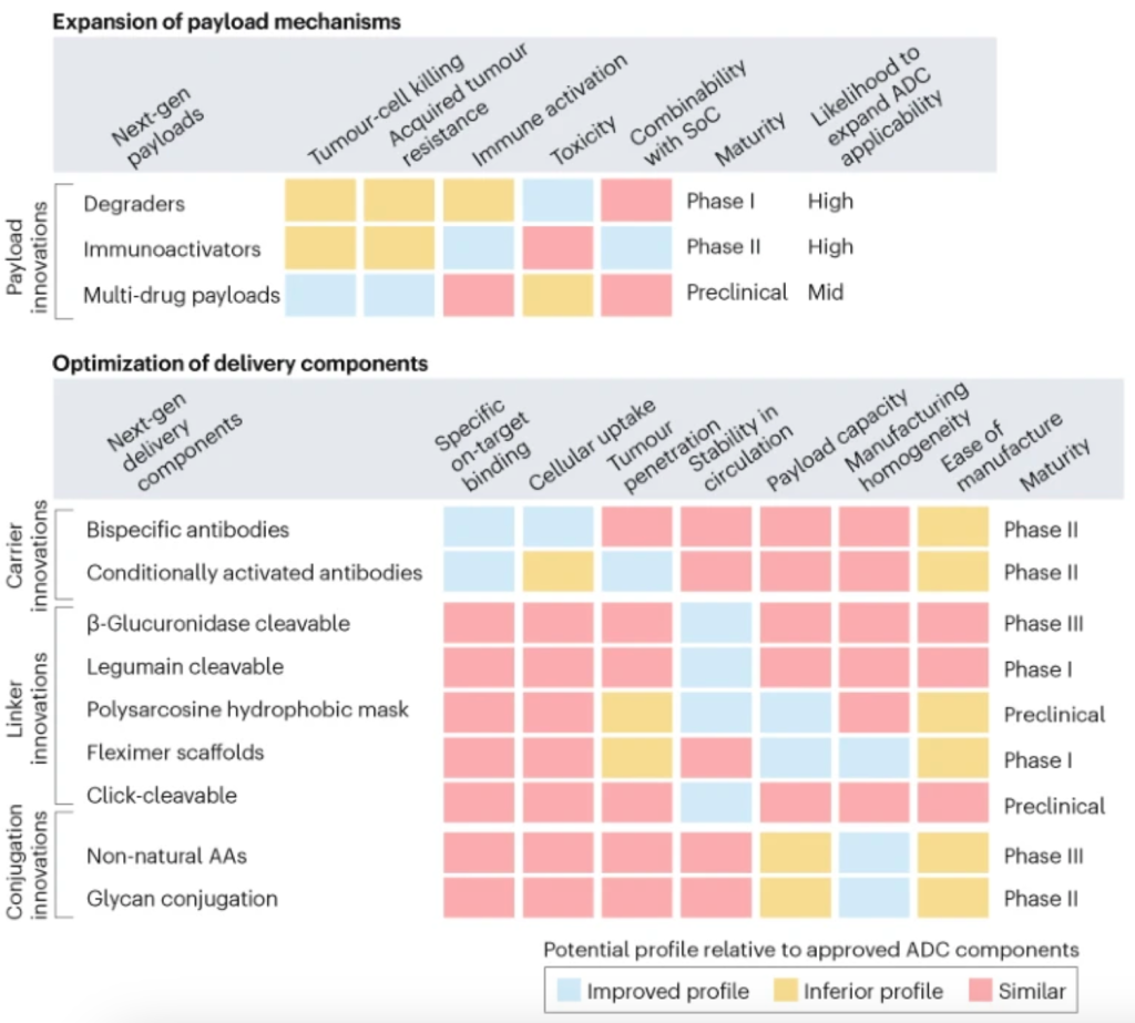
Microexon alternative splicing

Microexons are small sized (≤51 bp) exons which undergo extensive alternative splicing in neurons, microglia, embryonic stem cells, and cancer cells, giving rise to cell type specific protein isoforms. Due to their small sizes, microexons provide a unique challenge for the splicing machinery. They frequently lack exon splicer enhancers/repressors and require specialized neighboring trans-regulatory and cis-regulatory elements bound by RNA binding proteins (RBPs) for their inclusion. The functional consequences of including microexons within mRNAs have been extensively documented in the central nervous system (CNS) and aberrations in their inclusion have been observed to lead to abnormal processes. Despite the increasing evidence for microexons impacting cellular physiology within CNS, mechanistic details illustrating their functional importance in diseases of the CNS is still limited.

PTBP, known as hnRNPI, binds the polypyrimidine-rich region (U/CUCUCU) within introns and affects neuronal AS (Gil et al., 1991; Patton et al., 1991; Zheng, 2020). PTBPs has been extensively shown to be involved in AS of microexons in neurons during different contexts (Black, 1992; Chan & Black, 1995; Markovtsov et al., 2000). Neural progenitor cells abundantly express PTBP1 and during neurogenesis the expression of PTBP1 decreases, while the expression of its paralog PTBP2 increases (Chan & Black, 1997; Makeyev et al., 2007; Spellman et al., 2007). As a result, the synchronization of PTBP paralogs is critical for neuronal development and the switching of neuronal programs. The CLIP and RNA-seq data reveal that PTBP1 regulates microexon AS by binding upstream of the microexon (Y. I. Li et al., 2015). In the Neuro2A mouse neuroblastoma cell line, PTBP1-depletion caused microexon inclusion (~94%), whereas only 8% showed exclusion, inferring that PTBP1 is a repressor of microexon inclusion (Y. I. Li et al., 2015). This is in line with previous work from Black’s lab, where PTBP1 represses the N1 microexon inclusion of c-src mRNA in non-neuronal cells (Black, 1992; Chan & Black, 1997; Min et al., 1995). Likewise, PTBP1-depletion caused microexon skipping within the eIF4G transcript in neurons (Gonatopoulos-Pournatzis et al., 2020). Another study demonstrated that in the neural progenitor cell, microexon 5 of BAK1 transcript is skipped and this is promoted by the PTBP1 binding to the intronic region proximity to the 3′ splice site of the microexon. However, as neural progenitor cells differentiate to neurons, PTBP1 expression decreases, allowing the microexon to be included in the BAK1 transcript triggering the loss of BAK1 protein and enhancing neuronal survival (Lin et al., 2020). Therefore, PTBP1 is a microexon AS regulator playing crucial roles in neurons.

The physiological consequences or the causality of mis-spliced microexons has not been functionally examined. Possible ways to solve such a conundrum include performing gene editing with e.g., CRISPR/Cas9 to precisely remove individual microexons or flanking RNA elements and examine the functional outcomes (Du et al., 2020; Yuan et al., 2018). This approach will improve our comprehension of different small GTPase protein isoforms in regulating cellular physiology and CNS function. The observation of microexons AS in autism spectrum disorders is the beginning of mining these splicing events, especially of small GTPase regulators in CNS disorders at large, and determining whether microexon AS defects are a common feature of other disorders.

Source: PMID 34155820

A glimpse at the glycoRNA world

RNA modifications, discovered decades ago, have important biological functions. The most functionally validated modification is the 5′ m7G cap of mRNAs that controls canonical translation (Wei et al., 1975). A wide variety of modifications are present in tRNAs that affect its folding as well as translation. In the past decade, there has been an explosion in the number of known RNA modifications, particularly in mRNAs, and the discovery of their biological roles has spawned the important field of epitranscriptomics (Nachtergaele and He, 2018). In this issue in Cell, Flynn et al., link glycol- and RNA biology with the discovery of a new biopolymer, glycoRNA, a class of RNAs that are glycosylated with sialic acids and fucose (Flynn et al., 2021)

Paradigm shifts often require the development and implementation of tools to dissect and study dark spaces in biology. One approach for isolating, analyzing, and imaging glycosylated biomolecules is to co-opt the cellular biosynthesis of glycans by providing N-azidoacetylmannosamine (Ac4ManNAz), allowing researchers to label sialic-acid-containing glycans with a bioorthogonal handle (Baskin et al., 2007; Saxon et al., 2002). Although used broadly to detect glycosylated proteins, Flynn et al. report the first use of this approach to probe glycosylated RNAs. Indeed, glycoRNAs were detected in various human cell lines and in mouse liver and spleen tissues.

Although many modified mRNAs are found in the epitranscriptome, glycosylated mRNAs were not found. Rather, glycoRNAs are small nuclear (sn)RNAs, ribosomal (r)RNAs, small nucleolar (sno)RNAs, tRNAs, and Y RNAs, the latter of which comprise the greatest percentage of glycosylated RNA species. Further, fractionation and immunohistochemical imaging studies revealed that glycoRNAs are mainly associated with the cell surface, experimentally supported by their loss from the cell surface upon treatment with an enzyme the cleaves sialic acid (Figure 1). That Y RNAs are glycosylated is particularly interesting. Small, conserved RNAs that form ribonucleoprotein complexes, Y RNAs are known antigens associated with autoimmune diseases such as lupus. Because of this disease association and conservation, a series of rigorous experiments were completed to validate Y RNA glycosylation. In particular, CRISPR-Cas9 knockout of Y RNAs in HEK293T cells, which did not affect cell growth as expected from previous studies, ablated Ac4ManNAz-labeling of cells.

Next, the authors investigated whether the same biosynthetic machinery that produces N- and O-linked glycans used to glycosylate proteins also glycosylate RNA. They employed both genetic and pharmacological inhibition approaches. In cells where the glycan biosynthetic machinery is impaired by genetic manipulation, production of glycoRNA is impaired, which can be reversed by supplementation with exogenous glycan. Pharmacological inhibition of oligosaccharyltransferase also diminishes production of glycoRNA. Each study supports that the glycan biosynthetic machinery also produces cellular glycoRNA.

Expression of glycoRNA on the cell surface suggests it may play a role in signaling. It has been assumed that all cell-surface interactions of sialic acid binding-immunoglobulin lectin-type (Siglec) receptor family is due to its binding to glycolipids or glycoproteins. The Siglecs are the largest family of sialoside-binding proteins in humans, and they have important roles in various diseases, from cancers to autoimmune disorders to host-pathogen interactions. Flynn et al. show that two members of the Siglec family (−14 and −11) (Crocker et al., 2007) have interactions with the cell surface that are sensitive to RNase treatment, suggesting that glycoRNA mediate these interactions (Figure 1).

Armed with knowledge of the biopolymer, these rigorous and thorough studies lay the foundation to investigate the exact architecture and structure of glycoRNA; how the glycans are synthesized and incorporated into RNA, which RNAs are subject to glycosylation; and the regulation of its biosynthetic pathway. Most importantly, the precise biological functions of glycoRNAs can be determined. It was only a few decades ago that both RNA and glycans were an afterthought as direct players in human biology. Now that they have chemically joined forces, we should look forward to learning about how glycoRNAs affect biological processes!

References

J.M. Baskin, J.A. Prescher, S.T. Laughlin, N.J. Agard, P.V. Chang, I.A. Miller, A. Lo, J.A. Codelli, C.R. Bertozzi Copper-free click chemistry for dynamic in vivo imaging. Proc. Natl. Acad. Sci. USA, 104 (2007), pp. 16793-16797

P.R. Crocker, J.C. Paulson, A. Varki Siglecs and their roles in the immune system. Nat. Rev. Immunol., 7 (2007), pp. 255-266

R.A. Flynn, K. Pedram, S.A. Malaker, P.J. Batista, B.A.H. Smith, A.G. Johnson, B.M. George, K. Majzoub, P.W. Villalta, J.E. Carette, et al. Small RNAs are modified with N-glycans and displayed on the surface of living cells. Cell, 184 (2021), pp. 3109-3124

S. Nachtergaele, C. He Chemical modifications in the life of an mRNA transcript. Annu. Rev. Genet., 52 (2018), pp. 349-372

E. Saxon, S.J. Luchansky, H.C. Hang, C. Yu, S.C. Lee, C.R. Bertozzi. Investigating cellular metabolism of synthetic azidosugars with the Staudinger ligation J. Am. Chem. Soc., 124 (2002), pp. 14893-14902

C.M. Wei, A. Gershowitz, B. Moss. Methylated nucleotides block 5¢ terminus of HeLa cell messenger RNA Cell, 4 (1975), pp. 379-386

Library Preparation for Next-Generation Sequencing:Dealing with PCR Bias

From mapping the entire human genome to personalizing treatments based on specific mutations in an individual’s genome, we have come a long way! Next-generation sequencing (NGS), almost single- handedly, has enabled this gigantic leap in progress. Transforming the tedious chain termination method to a more automated and high-throughput sequencing by synthesis technique, NGS revolutionized the understanding of genetic variations and their implications. Apart from sequencing of fragments of DNA (DNA-Seq), sequencing of whole genomes (whole genome sequencing), exomes or transcriptomes (whole transcriptome sequencing), epigenomes (Methyl-Seq and ChIP-Seq) or even single cells (single cell sequencing) is now possible at a reasonable cost. While all these approaches have yielded valuable information, they suffer from an inherent problem of incomplete or misrepresentation of data, which consequently leads to misinterpretation of information. The predominant factor leading to misrepresentation of data is the bias prevalent in almost all steps of NGS sample preparation. In this article, we focus on PCR bias in NGS library preparation and highlight a few publications where solutions to this bias have been documented.

Bias during amplification of AT- and GC-rich regions

During NGS library preparation, DNA or RNA molecules are fragmented, ligated to adapters suitable for the particular sequencer used, size selected and amplified using PCR. Most of the enzymatic steps within library construction protocols introduce bias in sample composition. One of the most likely sources of bias is the PCR amplification step, which could yield uneven base composition due to the fact that amplification is not uniform among fragments. Samples with high GC or AT content are not amplified as efficiently, and when this inefficiency is amplified exponentially over several cycles in PCR, it leads to notable inaccuracies in sequencing results. To avoid this, special caution is needed in selecting DNA polymerases used for the amplification step. A comparative study published in Nature Methods assessing biases in PCR amplification during NGS library preparation1 assessed the efficiency of several DNA polymerases under different reaction conditions to amplify adapter-ligated fragments for Illumina sequencing. They tested several microbial genomes with differing GC content (from approximately 20% to 70%) for depth of coverage under different experimental conditions, such as standard amplification, with a qPCR formulation or with annealing and extension at 60oC. Their results stated that KAPA HiFi DNA polymerase was the optimal enzyme for NGS library amplification. Genomic coverage was also reported to be highly uniform using the KAPA HiFI DNA polymerase enzyme, and was very close to results obtained without PCR for all tested GC contents.1

Efficient amplification of AT-rich regions require low annealing temperatures, but this often results in misannealing and nonspecific amplification.2 Tetramethyleneammonium chloride (TMAC), a DNA-binding reagent, is often added in PCR reactions of samples with high AT content for increasing the melting temperature, and consequently, the thermostability of AT pairs. However, TMAC by itself could inhibit the polymerase activity of some enzymes. A study that explored optimal library preparation procedures for samples with high AT content tested several enzymes (Phusion, AccuPrime Taq HiFi, Platinum pfx, KAPA HiFi and KAPA2G) and found that among all of them, only KAPA HiFi and KAPA2G Robust were able to amplify the AT-rich locus efficiently in the presence of the TMAC additive.3 This study also confirmed that KAPA HiFi DNA polymerase amplified the AT-rich Plasmodium falciparum genome more uniformly and provided the best coverage compared to all the other enzymes and that its amplification and coverage depth were closer to that of PCR-free conditions.

PCR-free amplification could yield better read distribution and coverage compared to PCR methods, but would require large quantities of starting DNA material. Therefore, this method is not highly practical to use, especially when sample volume is limiting (such as in FFPE samples).

PCR bias during library preparation for RNA-Seq

RNA-Seq also faces several challenges during library preparation, such as removal of highly abundant ribosomal RNA and PCR bias during amplification of the adapter-ligated library. A publication that reviewed reported biases in DNA and RNA library preparation4 found that KAPA HiFi DNA polymerase performed better than most enzymes and suggests that KAPA HiFi is a better choice than traditional polymerases for the amplification step. Since the RNA-Seq workflow includes more steps to convert RNA to cDNA prior to library construction, reducing PCR bias could help alleviate bias introduced in the process.

Dealing with bias

Given the extreme complexity of the NGS library construction and sequencing process, bias is something that cannot be entirely eliminated. The best way to mitigate bias is to recognize where the possible sources are introduced and use the most optimal library construction reagents. There are some comparative studies and reviews with extensive analysis of the sources of bias in each step of library preparation.1,4 These studies have evaluated the performance of library preparation reagents under different conditions and have made recommendations. Therefore, instead of reinventing the wheel, you may be able to utilize the optimized protocols and reagents directly and fine-tune them for your specific applications. Some studies have focused on specific biases (for example coverage of genomes with extreme AT-rich regions) and have developed optimized protocols for them.3 Using these pre-optimized protocols and reagents documented and recommended in published work could save time, cost and effort.

References

Quail MA, Otto TD, Gu T et al. Optimal enzymes for amplifying sequencing libraries. Nat.Methods. (2012);9:10–11.
Chevet E, Lemaitre G and Katinka MD. Low concentrations of tetramethylammonium choloride increase yield and specificity of PCR. Nucleic Acids Res. 1995;23;16:3343-3344.
Oyola SO, Otto TD, Gu Y et al. Optimizing Illumina next-generation sequencing library preparation for extremely AT-biased genomes. BMC Genomics 2012;13:1
van Dijk EL, Jaszczyszyn Y, Thermes C. Library preparation methods for next-generation sequencing: Tone down the bias. Experimental Cell Res. 2014;322:12-20.

核酸提取试剂盒的原理

A Step-by-Step Guide to Nucleic Acid Extraction Kits
Step 1: Cell Lysis

Lysis formulas may vary depending on whether you want to extract DNA or RNA. Generally speaking, lysis buffers contain a high concentration of chaotropic salts. Chaotropes have two important roles in nucleic acid extraction:

They destabilize hydrogen bonds, van der Waals forces, and hydrophobic interactions, leading to destabilization of proteins, including nucleases;
They disrupt the association of nucleic acids with water, thereby providing optimal conditions for their transfer to silica.

Chaotropic salts include guanidine HCL, guanidine thiocyanate, urea, and lithium perchlorate.

In addition to chaotropes, a detergent is often present in the lysis buffer to aid protein solubilization and cell lysis.

Enzymes may also feature here, depending on the sample type. The broad-spectrum serine protease Proteinase K is very efficient in digesting proteins away from nucleic acid preparations. Proteinase K works best under protein denaturing conditions (i.e. in denaturing lysis buffer). Another popular enzyme here, lysozyme, does not work under denaturing conditions and will be most active before the addition of denaturing salts.

Bear in mind that lysis for plasmid isolation is very different from lysis for RNA or genomic DNA extraction because plasmids must be separated from genomic DNA first. The addition of chaotropes will release all types of DNA at once, losing the ability to differentiate small circular DNA from high molecular weight chromosomes. Therefore, in plasmid preps, the chaotropes are not added until after cell lysis. For additional reading, check out these great articles on alkaline lysis and plasmid and genomic DNA extraction.


Step 2: Purification – Binding Nucleic Acids to the Column

As discussed above, chaotropic salts are critical for lysis and binding to the column. The addition of ethanol (or sometimes isopropanol) will further enhance and influence the binding of nucleic acids to the silica.

Note that the percentage and volume of ethanol used are important. Too much ethanol will bring down degraded material and small species that will influence absorbance at 260 nm (A260 readings). On the other hand, too little ethanol may impede the washing of the salt from the membrane.

Fortunately, the amount of ethanol added will be optimal for the nucleic acid extraction kit you are using. However, if you suspect that degraded DNA is inflating your A260 readings, you can consider re-optimizing the ethanol concentration.

Another useful tip is to save the flow-through and precipitate it to see if you can find your lost material. If you used an SDS-containing detergent for lysis, try using NaCl as a precipitant to avoid detergent contamination of your nucleic acids.


Step 3: Washing

After centrifuging your lysate through the silica membrane the desired nucleic acids should be bound to the column and impurities such as protein and polysaccharides should be in the flow-through. However, the membrane will contain protein and salt residues. At this point, plant samples will likely contain polysaccharides and pigments, while for blood samples, the membrane may be slightly brown or yellow in color. The wash steps remove such impurities.

There are typically two wash steps, although this varies depending on sample type. The first wash will often include a low concentration of chaotropic salts to remove residual proteins and pigments. This is always followed by an ethanol wash to remove the salts. If the sample didn’t contain a lot of protein starting out (e.g., plasmid preps or PCR clean-ups), an ethanol wash is sufficient.

Removal of the chaotropic salts is crucial to getting high yields and purity. Some kits actually recommend two ethanol washes. Residual salt will impede the elution of nucleic acid, resulting in poor yield, high A230 readings, and thus low A260/230 ratios.


Step 4: Dry Spin for Ethanol-free DNA and RNA

Most protocols include a centrifugation step after washing to dry the column of residual ethanol, and this step is essential for a clean eluent. Subsequent addition of 10 mM Tris buffer or water to the membrane will hydrate the nucleic acids, thus eluting them from the membrane. Residual ethanol on the membrane at this point will prevent full hydration and elution of nucleic acids.

You will not be able to see ethanol on a spectrophotometer, but a good indicator of its presence is samples that will not sink into the wells of an agarose gel, even in the presence of loading dye. Another indicator of ethanol contamination is samples that don’t freeze at -20°C.


Step 5: The Final Frontier – Elution

The final step in the DNA extraction protocol is the release of pure DNA or RNA from the silica.

For DNA extraction, 10 mM Tris at pH 8-9 is typically used. DNA is more stable at a slightly basic pH and will dissolve faster in a buffer than water. This is true even for DNA pellets. Water tends to have a lower pH of 4-5, and high molecular weight DNA may not completely rehydrate in the short time used for elution. For maximal DNA elution, allow the buffer to stand in the membrane for a few minutes before centrifugation. For applications requiring intact high molecular weight DNA, such as long-range sequencing and long-read sequencing, elution buffer is the best choice.

RNA, however, can tolerate a slightly acidic pH and dissolves readily in water, making this the preferred diluent.


What Can Go Wrong with Nucleic Acid Extraction Kits?


Low Yields

If you experience lower yields than you expect, there are many factors to consider. It is often a lysis issue, with incomplete lysis being a major cause of low yields. Incorrect binding conditions is another possibility. Make sure to use fresh high-quality ethanol (100%, 200 proof) to dilute buffers and for the binding step. Low-quality ethanol or old stocks may have taken up water, skewing the actual working concentration. Remember that if you make your wash buffer incorrectly, you may be washing away your extracted DNA or RNA!
Low Purity

If the extract is contaminated with protein, you may have started with an excess sample, increasing the risk of incomplete solubilization. If the extracts have poor A260/230 ratios the issue is usually residual salt after binding or inadequate washing. Be sure to use the highest quality ethanol to prepare wash buffers and if the problem continues, perform an additional wash step.


Impurities

Environmental samples are especially prone to impurities because humic substances solubilize easily during extraction. Such substances often behave similarly to DNA during the extraction process and are difficult to remove from the silica column. For samples prone to impurities, specialized techniques exist to remove interfering protein and humics prior to column binding.
Degradation

This is a greater concern for RNA than DNA extraction and you can find specific advice on troubleshooting RNA extraction here. RNA degradation often occurs due to improper sample storage or inefficient lysis, assuming of course samples are eluted with RNase-free water. For DNA extractions, degradation is not a huge issue if PCR is the desired application, but if you were hoping for intact high molecular weight DNA for long-range sequencing applications you should ensure to not be too harsh when lysing your sample!


PCR Clean-up Special Considerations

PCR cleanup isn’t a DNA extraction technique per se, but it is worth a mention here. Typically, PCR products are cleaned up by adding 3-5 volumes of salt per volume of the PCR reaction, followed by centrifugation of the mixture through a spin column. Although a failed clean-up is often caused by an unsuccessful PCR, it is worth saving your flow-through after column binding. If a strong PCR band didn’t make it through the column, chances are it is in the flow-through. You can always rescue it and clean it up again.
Go Forth and Use Your Nucleic Acid Extraction Kits with Confidence

As scientists, we often want to be able to troubleshoot without asking for outside help. This article should clarify some of the science around silica spin filter technology in many nucleic acid extraction kits allowing you to troubleshoot in no time. If all else fails, you will have done your homework by the time you call for technical support, and you should reach a resolution much faster, even if that is a free replacement DNA extraction kit!

Do you have any comments or questions about how nucleic acid extraction kits work? Leave a comment below.

家庭网络计划的购买:注意隐藏的费用

合约可能的隐藏费用

数据机费用Modem Rental Fee

有些业者供月租服务,通常是$6-$10/月。但老实说,一台Modem约$60至$100上下,如果你很确定会长期使用网络,自己买一台还比较省钱!(别忘了购买与网络业者兼容的Modem机型)

安装费Installation Fee

虽然你跟业者订购了网络服务,但可不代表他就会免费帮你安装!在你跟技术人员约时间作业前,别忘了先问问是否有安装费用会产生,这费用可是从$25到$100不等。说不定你身边就有亲友(或你自己)会安装Router,直接省下一笔。

服务启动手续费Activation Fee

又是一条业者挖钱的好名目,随便又是$50飞走。在订购服务前,别忘了先跟业者确认是否有这笔费用。此外,如果你当初不是电视、网络等服务一次同时订购,分次订购业者也是会跟你分次收取启动手续费。你可以打电话去尝试将这笔费用移除。

提早取消服务费Early-Termination Fee

部分业者会针对有签合约者提早取消服务收取一笔解约费用,合约中小字别忘了读仔细。

到府服务费Service Call Fee

不管是任何大小问题,只要有技术人员前往你家就有可能会被酌收一笔到府服务费用,所以下次预约到府服务前,别忘了先问清楚会不会有费用产生,如果有,也别忘了了解此项费用的涵盖内容为何,例如是因为业者机器问题导致你的网络无法使用而要求技工前往维修,那你是否还要付这笔费用。

来自: https://www.extrabux.com/chs/guide/4252214

single-cell RNA-seq: a collection

The motivation to collect resource from the web, is the problem occurring in single-cell RNA seq: cells are lost after cell-death removal kit.

Why happens? how to solve the problem?

  1. The Single-Cell Preparation Guide https://genome.duke.edu/sites/default/files/Single-Cell-Prep-Guide.pdf
  2. Removal of Dead Cells from Single Cell Suspensions for Single Cell RNA Sequencing https://assets.ctfassets.net/an68im79xiti/i1zxeDHukSQKEs2imsKYk/6a8429045a0666f0f4f1a4f908234f0b/CG000093_SamplePrepDemonstratedProtocol_-_DeadCellRemoval_RevA.pdf
  3. Why is my cell recovery low after using the Dead Cell Removal protocol (CG000093)? https://kb.10xgenomics.com/hc/en-us/articles/360044580971-Why-is-my-cell-recovery-low-after-using-the-Dead-Cell-Removal-protocol-CG000093-
  4. An optimized workflow for single-cell transcriptomics and repertoire profiling of purified lymphocytes from clinical samples https://www.nature.com/articles/s41598-020-58939-y
    5.Cell fixation and preservation for droplet-based single-cell transcriptomics https://bmcbiol.biomedcentral.com/articles/10.1186/s12915-017-0383-5
  5. Book_Single Cell Methods and PROTOCOLS http://shaleklab.com/wp-content/uploads/2019/05/2019_Book_SingleCellMethods.pdf?
  6. Droplet-based single cell RNAseq tools: a practical guide† https://pubs.rsc.org/en/content/articlelanding/2019/lc/c8lc01239c#!divAbstract
  7. Complete Guide to Understanding Single-Cell RNA-Seq https://www.activemotif.com/blog-single-cell-rna-seq
  8. What is the best way to separate the viable cells from the dead cells in suspension culture? https://www.researchgate.net/post/What_is_the_best_way_to_separate_the_viable_cells_from_the_dead_cells_in_suspension_culture
  9. A reliable strategy for single-cell RNA sequencing analysis using cryoconserved primary cortical cells https://www.sciencedirect.com/science/article/pii/S0165027020303836
  10. Sampling time-dependent artifacts in single-cell genomics studies https://www.ncbi.nlm.nih.gov/pmc/articles/PMC7212672/
  11. Fresh Frozen Human Peripheral Blood Mononuclear Cells for Single Cell RNA Sequencing: https://assets.ctfassets.net/an68im79xiti/6xwl38JmJc32Y1dgKq6dSt/32c8fc43de6dd213717b34edc44c53c3/CG00039_Demonstrated_Protocol_FreshFrozenHumanPBMCs_RevD.pdf
  12. Single Cell Set-Up: Sample Preparation Tips https://www.biocompare.com/Bench-Tips/345311-Single-Cell-Set-Up-Sample-Preparation-Tips/
  13. Quality Control of RNA-Seq Experiments: https://link.springer.com/protocol/10.1007/978-1-4939-2291-8_8
  14. Best Practices for Illumina Library Preparation https://currentprotocols.onlinelibrary.wiley.com/doi/10.1002/cphg.86

马桶杂音的解决

家里马桶冲水后,一直响个不停。去年发生一次,后来买了一个配件解决了。

今年同样的问题再次发生,但是记不清去年怎么解决的了。只好开车去lowes转了一圈,买回来Toilet Fill Valve Seal

https://www.lowes.com/pd/Fluidmaster-Rubber-Toilet-Hardware-Kit-For-Fluidmater-400A/1000106791

折腾了半天,关键是上面的水管头部怎样拧下来。看了官方视频,才知道是提起浮标杆,然后摁压盖子,逆时针旋转1/8,就下来了。里面的一个黑色塑料片是耗材,每隔一段时间就要更换:

操作视频:https://www.youtube.com/watch?v=oD0HETCc-OQ

RNA-seq Sample Guidelines

The key to successfully performing RNA-seq experiments is to provide the core facility with total RNA of sufficient quality and quantity to produce a library for sequencing. The quality of the initial samples is by far the single-most important factor in the whole process.

Tissue and Cell Isolation
Investigators need to carefully choose their methods of tissue and cell isolation, as these methods directly impact the quality and quantity of RNA that is subsequently obtained. If possible, total RNA purification should immediately follow tissue/cell isolation to prevent alterations in the transcript profile. However, in some cases, immediate RNA purification is not possible. If tissues/cells must be stored prior to RNA isolation then the use of products, such as RNALater from Qiagen or similar reagents, is recommended.

The core facility strongly encourages pilot projects to confirm that the chosen methods will reproducibly create sufficient quantities of cells/tissues to ultimately yield the required amount of high-quality RNA. Once an isolation protocol or a storage and isolation protocol is established, it is important that all of the samples collected for a given project be collected with this same protocol. Variance in these techniques may result in differences in the transcript profile. These differences may later be misidentified as changes due to experimental treatment, while in reality they are artifacts of varying isolation and storage methodology.

RNA Isolation
The facility does not perform total RNA purification. We require a minimum of 500 ng of total RNA for QC and library preparation for Illumina sequencing. A number of well-established commercial kits and protocols exist for a variety of species and tissue/cell types. Investigators carefully need to determine the most appropriate methods for their tissue/cell type. The core facility strongly encourages pilot projects to confirm that the chosen method will reproducibly create sufficient quantities of high-quality total RNA from the tissue/cell of interest. Given the tremendous variety of tissue/cell types, it is difficult for us to give specific recommendations. For anyone not sure of what product to choose, we strongly encourage you to examine the products from Qiagen and Ambion (LifeTech) as a starting point. These companies have a large variety of products with decision charts to help you in choosing the right one. In addition, if you need advice that is more detailed, there are people in tech support at these companies who truly are experts in RNA isolation and have a broad experience in helping customers isolate RNA from every conceivable species/tissue/cell. We however, do have a few general recommendations regarding RNA isolation techniques based upon our experience.

We do not recommend the use of Trizol alone for total RNA isolation, as the use of Trizol often results in samples that are contaminated by proteins and organics which can inhibit the library making process. We do recommend products like RNeasy, which is a column-based purification method producing very pure preparations of total RNA. Many of our customers have found though that they get superior yields with Trizol so they perform an initial isolation with Trizol followed by a further cleanup using the RNeasy kit. We have observed that this results in very pure RNA and this method has been used successfully for RealTime PCR, microarrays, and RNA-Seq.

Accurate Determination of RNA Concentration and Purity
RNA concentration is routinely determined by measuring absorbance at 260 nm. However, it should be noted that all nucleic acids have a peak absorbance at approximately 250 -260 nm; this includes RNA, DNA, and free nucleotides. If your RNA preparation contains contaminating DNA or free nucleotides it will affect your ability to determine accurately the RNA concentration in your sample with a spectrophotometer.

RNA purity is determined by measuring the 260/280 and 260/230 ratios using a spectrophotometer. Excessive absorbance at 280 indicates the presence of protein in your sample while excessive absorbance at 230 may indicate the presence of residual phenol in your sample. Ideally, the 260/280 ratio for RNA should be approximately 2.0 and the 260/230 ration should be 2.0-2.2.These ratios can be affected by pH so we are comfortable with all ratios over approximately 1.8. Ratios that differ significantly from that measure should undergo further purification.

We recommend the use of a NanoDrop to determine RNA purity and for an initial estimate of RNA concentration. This device is easy to use and less prone to error than traditional spectrophotometers because sample dilution is usually unnecessary and pipetting errors have no effect on correct determination of concentration. All facility customers may use the one we provide in 411 Chandlee, free of charge, but please bring your own pipettor and tips. This instrument is calibrated regularly so that customers may be assured it provides an accurate measurement.

We regularly see sample concentrations reported to us that are in wide variance from the true concentration measured by our facility with calibrated a NanoDrop or other techniques such as Bioanalzyer or Qubit. If you are using your own spectrophotometer please check that it is calibrated by measuring the concentration of commercially obtained standards, use calibrated pipettors, and be careful in calculating and performing your dilutions.

RNA Sample Quality
In addition to concentration and purity, it is essential to determine the quality of RNA samples prior to library preparation for RNA-Seq, to ensure that differential degradation of samples is not mistaken for differential expression. The quality of an RNA sample (its level of degradation) can’t be determined using the NanoDrop. Sample quality is determined using an Agilent Bioanalzyer. The Bioanalyzer will produce an RNA Integrity Number, or RIN, which is an objective measure of RNA quality. RIN scores vary from 1-10, with 10 being the highest quality samples showing the least degradation.

We not only like to see high RIN scores (7-10), but we also like to see a reasonably narrow range of the scores within a set of samples, which is typically 1-1.5. We recommend re-isolation of samples that have low RIN (6 or below) or are large outliers from the average RIN of a group of samples.

Ideally we would like 5 ul of total RNA at a concentration of 100 -200ng/ul to perform Bioanalzyer analysis and to confirm sample concentration and purity. Once the total RNA samples have passed these quality measures, the samples can be used for library preparation which requires approximately 300 ng of total RNA.

If the customer plans on performing rRNA depletion, we recommend checking each total RNA sample prior to depletion to confirm quality. Following depletion, the samples should once again be assessed on the Bioanalzyer to determine the success of the depletion.

Summary
High sample quality is essential for successful RNA-Seq experiments.
Customers are responsible for total RNA isolation.
We encourage customers to perform pilot projects to determine the best tissue/cell isolation technique and RNA purification technique for their sample type.
Once a tissue/cell isolation technique and RNA purification technique has been established it should be adhered to for all samples in a project.
Determine total RNA sample purity and estimate sample concentration with a NanoDrop. The 260/280 and 260/230 ratios need to be greater than 1.8. Enough total RNA must be isolated to provide us with 500 ng for sequencing on the NextSeq or MiSeq.
Determine total RNA quality prior to library construction by having the Genomics Core Facility assess the sample using the Agilent Bioanalzyer. RIN of 7-10 and ranges of RIN from 1-1.5 for a group of samples are preferred.

Many types of RNA-seq require RNA samples of high integrity and high chemical purity – please see the sample requirements. If the tissue or cell samples are handled correctly (e.g. flash frozen and stored at -80C) standard spin column RNA extraction kits will yield RNA samples perfectly suitable for RNA-seq. Please note that samples destined for miRNA or small RNA studies need to be isolated with protocols specifically designed to retain the small molecules (please see below). Standard RNA isolation protocols will lead to the loss and sequence-specific selection of small RNA molecules. RNA samples should always be DNA-free. Nanodrop readings are more or less useless to determine RNA sample concentrations – please use fluorometric quantification instead (e.g. Qubit or Quantus instruments). The Nanodrop readings should be used to assess sample purity.

Avoiding Batch-Effects:
Both sample storage conditions and details of the RNA-isolation protocols are well-known to introduce technical variations into RNA-seq data. Because of this, it is recommended to:

Isolate the RNA-samples in one batch.
If RNA-isolations need to be carried out in several batches, they should be carried out by the same person using the same batch of reagents
If RNA-isolations need to be carried out in several batches, the samples should be randomized between the RNA isolation batches (worth discussing with a statistician or the Bioinformatics Core).

中国研究生在美求职及时机

本文作者“子陵在听歌”,系作者在新浪微博原创文章。

收到了不少私信,问生物医学PhD毕业后应该选择工业界还是学术界。其实我早就想就这个话题写一篇文章,但并不是想指导或教育谁,而是告诉大家许多过去的教训。就像我读PhD的第一年有很多失败教训,比如什么课不该选,什么老板实验室不能去做rotation,以后中国学弟学妹来了我都告诉他们这些,确实让一些人少走了很多弯路。正巧这一周我和我三个研究生博士最好的朋友(他们都在博士后最后阶段)聊天,发现了我们有很多共识。这里有几点分享一下:

  1. 一定要利用好OPT。OPT的工卡(EAD)是绿卡EAD之前最好的一个工作签证,3年时间可以随意换雇主,有5个月grace period,因此可以抽3次H1b。这3年时间宝贵,千万不要浪费(比如OPT挂靠在博士实验室一年半帮博士老板打工)。尤其是有志博士毕业直接去工业界工作的同学,千万不要去做学术界博士后浪费时间,而一定充分利用这3年找到工业界工作或做工业界博士后,最后都可以找到最好的工业界工作机会并抽中H1b。
  2. 我觉得大部分人博士毕业都没有明确目标,并不知道博士毕业该去学术界还是工业界。这是正常的。所以绝大多数人都倾向于做学术界博士后以观望,这也是我当时的心态。我PhD入学的时候,系主任在orientation的时候就说过,差不多每50个生物PhD学生最后只有3-5个能拿到tenured faculty,这还包括很多社区大学职位或teaching faculty职位。而能够入选PhD项目的人智商和努力程度都没什么问题,所以我朋友和我都觉得最后能不能拿到faculty很大程度是运气和机缘。但是,如果你确实还有想做faculty留在学术界的想法,博士期间一定要牢牢掌握好基础知识、基本技能,争取发两篇以solid实验为基础的本领域认可杂志的一作文章,这是找好博士后从而当faculty的敲门砖。博士毕业前半年好好networking,利用博士老板的资源找个好postdoc。我认识绝大多数找到faculty的都是博士后在大牛实验室完成的。每个领域的大牛定义不一样,比如我熟悉的HIV领域很少有院士和HHMI,但是我觉得最可以关注的一个指标就是这个实验室能否培养中国faculty。如果这个实验室近5年不断能有领域内顶级论文发表,一直有中国postdoc能够找到faculty,那么这个实验室是很好的通往学术界的选择。还有实验室氛围不toxic也十分关键。
  3. Postdoc申请有许多trick,其中很重要的一点就是H1b sponsorship,这点不妨直接给学校的国际留学生处打电话询问。如果OPT 3年内能拿到H1b,确实省去了很多时间和麻烦,从而可以尽快开始NIW和EB1的申请。但签证不应该成为决定去不去做博士后的因素,在这个实验室工作的前途才应该是决定因素。
  4. Postdoc尽量选择3个大城市纽约、波士顿和湾区,这是因为实验室多,万一想转组机会多,将来博士后结束如果想去工业界机会也多,尤其是波士顿和湾区。美国公认3个做博士后最集中的地方,分别是波士顿哈佛医学院及附属医院-哈佛本部-MIT及附属研究所;纽约RU-Sloan Kettering-Weill Cornell三校及哥大;旧金山湾区UCSF-Stanford-UC Berkeley。在这些地方博士后有不少福利,尤其是后两者,有打折宿舍等。而且这些地方产学研交织,校友网络质量极高,很容易为将来找学术界和工业界工作奠定基础。
  5. 博士后如果想留在学术界,时间节点确实很重要。4年之内最好有文章产出并申请K99/R00,如果有绿卡还可以申请K22。因此在前3.5年如果看不到文章产出的可能性,课题需要重要调整甚至换方向或者转组,可以考虑是不是要继续做postdoc。这个时候虽然因为身份原因,找工业界工作难度上升,但是可以开始networking和收集信息,联系之前去工业界工作的同学同事进行informational interview。因为这时再在学术界逗留,有可能会出现所谓“千老”的情况。当然,我也认识做了6-8年postdoc在最后两年忽然爆发发表文章从而找到faculty的情况,因此“时间点”也不能一概而论。
  6. 在有OPT或者绿卡的情况下,找到工业界工作的周期是3-6个月。这里with all due respect,但是中国人非常热衷去“大厂”,觉得这样有面子。我专门找了两个美国career coach,每个季度付费和他们交流,他们都非常不认同这种想法。工作一定要找合适的,而不是找有面子的。如果你喜欢做原创研发,大部分大药厂并没有这种机会;相反,小公司有更好的创新平台。除此以外,大公司的福利可能更好,但在波士顿和湾区小公司可能会给entry level scientist更高的title和工资,因此很多情况可能适合于刚刚毕业的PhD或者刚刚做完postdoc的人。找工业界工作最大的两个诀窍就是改好简历并用好Linkedin。
  7. 关于改简历,学术界用CV,工业界用resume,resume是求职敲门砖,力求简洁,应该在2页内。初次找工业界工作的人往往都会犯一个错误,就是在resume上写入很多的内容。因此申请第一个工作的时候,不妨花$100-200找个resume写手帮你写个resume并写一个cover letter模板。但resume的作用核心是通过网络工作申请系统的AI审查,因此,每一个工作申请的resume都应该根据工作要求重新编辑,强调你能掌握这个工作所需要的核心技术。
  8. 关于LinkedIn,这是求职最重要的社交媒体平台。多关注别人并加好友,尤其加各个你感兴趣公司的HR,他们经常会post手头的职位,还会转发需要招聘的hiring manager的求聘post,并鼓励你直接联系他们。另外LinkedIn可以帮你找到博士后或者博士学校的校友,这个共性可以作为你在LinkedIn加对方好友时发信息的基础。而informational interview是去你想去公司的敲门砖之一,因此,在LinkedIn上一旦加了好友,可以邀请对方给你个15分钟informational interview,以了解你感兴趣的公司的各种信息,尤其是有没有职位开放,甚至有没有可能内推。
  9. 工业界工作面试一般流程是网络系统申请,HR初筛,hiring manager电话面试,panel interview(终面)。每一步之间大约1-7天不等,但一旦过了2周,很大可能是没有被选中。面试有太多注意事项,但我觉得很重要的一点就是要反复读自己的简历和该职位的招聘信息。面试你的人也基本都是拿着你的简历提问题的。在panel interview后,如果公司对你感兴趣,会1-2周内很快给你发offer。一旦最初的offer发下来,这个时候可以讨价还价。Glassdoor网站有各个地区各个level的职位的平均工资标准,可以以此为market value进行5-10k的讨价还价。但大部分情况,公司很难给你上涨超过他们最初offer的10k以上。一般来讲,工资是跟title挂钩的。除此以外,福利也比较重要,如PTO,401k贡献比例,股票或者期权,这些其实都能转化成钱。另外,glassdoor还有各个公司员工对这个公司的review,可以以此看出这个公司的福利待遇和企业文化是不是合适你,以此作为判断拿不拿这个offer的标准。
  10. 许多人认为博士后做得不顺利,但是由于是OPT最后一年或者J1身份因此无法找工业界工作。其实这种情况只是“难”,但不是完全没办法。比如很多公司就可以赞助O1签证,因此不需要J1 waiver。这时候其实只需要做好一个心理准备就是不会在3-6个月找到工业界工作,而可能要12-18个月。这时需要做的,是突出你的transferable skills并坚持做informational interview,比如一个公司招聘会一种技术的科学家,你就要突出你对这一技术的熟练掌握程度,比如突出发表的文章或者会议摘要等,并可以在LinkedIn上联系该公司该项目的科学家。找工业界工作就是有志者事竟成,只要下定决定,最后都会找到。

Extension service: The go-to place for growing concerns

With cars whizzing by on a nearby highway, the Cornell Cooperative Extension marked the spot where it began 100 years ago when a farmer set out in a horse and buggy to help local growers. The farmer, John Barron, was recently honored with a stone marker next to a bank on a former field in Binghamton, NY. And while the landscape has changed dramatically, the mission of the extension program, now in every state, has not.

Today there are 2,900 extension offices across the country that not only help farmers raise crops but tell homeowners how to get greener grass and identify the pests in their yards. Overseen by the U.S. Department of Agriculture and administered by the state land grant colleges and universities, the extension service may seem a bit retro. That is until you need a cure for crab grass or find telltale signs of a varmint in your yard.

Every state has a cooperative extension system. To find the office closest to you check the map on the USDA’s website. In New York alone, 10 million people take advantage of information shared by the extension service on everything from growing fruit and vegetables to finding a farmer’s market.

The Cornell Cooperative Extension, for example, has a month-by-month guide to lawn and yard care. Here’s what they recommend for April. (If you live in a warmer region, check your state’s extension site for advice.)

Mowing. Begin when turf is 4 to 4 1/2 inches tall. Set mower height to 3 inches. Remove only one-third of the overall grass height with each mowing.
Fertilizing. Don’t fertilize yet if your lawn looked good after winter and/or you fertilized in late fall. Excess fertilizer in early spring promotes top growth at the expense of root growth. Deeper roots are more resistant to pests and drought.
Seeding. Seed only if you have a thin, weak lawn or bare spots. Depending on the species, grass will germinate when soil temperatures reach 45 to 55° F at the 2-inch depth.
Irrigating. Spring rains should be sufficient.
Monitoring soil temperatures. Insert a thermometer to 2 inches and wait 5 minutes.
Scouting. Search for large grubs, but do not apply insecticides because they will not be effective.

https://www.consumerreports.org/cro/news/2011/03/extension-service-the-go-to-place-for-growing-concerns/index.htm

https://www.consumerreports.org/cro/news/2012/10/husqvarna-introduces-a-tool-free-way-to-change-mower-blades/index.htm

https://www.consumerreports.org/cro/news/2008/03/tip-of-the-day-get-your-lawn-mower-or-tractor-in-shape-for-spring/index.htm

https://www.consumerreports.org/cro/news/2007/07/tip-of-the-day-sharpen-your-mower-blade/index.htm

https://www.consumerreports.org/lawn-mowers-and-tractors/how-to-get-your-lawn-mower-ready-for-spring/

https://www.consumerreports.org/lawn-care/a-cut-above-how-to-get-the-perfect-lawn/

OUTSTANDING QUESTIONS FOT TDP43

TDP-43 regulates hundreds of transcripts. Do a small number of these target genes account for disease pathogenesis and progression, or are TDP-43 proteinopathies the result of many modest molecular ‘paper cuts’ collectively summing to major dysfunctions?

Can restoration of a small number of TDP-43 target RNAs (e.g., STMN2) serve as a clinical strategy for TDP-43 proteinopathies, or do therapeutic approaches need to focus on pathways upstream of TDP-43 to restore a broader set of transcripts?

More than 50 mutations associated with disease have been identified in TARDBP . What are the consequences of these mutations on TDP-43 function, and do they lead to distinct or common defects?

The ability of TDP-43 to phase separate into biomolecular condensates is well established, but how is this process regulated in response to cellular stresses? Are there signal transduction cascades that regulate the ability of TDP-43 to specifically respond to these insults? If so, what are they?

TDP-43 pathology has also been observed in astrocytes. Does TDP-43 pathology in non-neuronal cell types also lead to significant alterations in RNA metabolism? Does pathology in non-neuronal cells contribute to disease onset or progression?

Reduced STMN2 expression has been observed in other TDP-43 proteinopathies. Is the mechanism behind reduced STMN2 expression also the consequence of premature polyadenylation and inclusion of a cryptic exon, or is it the result of neuronal loss?

Disrupted nuclear import-export in neurodegeneration

The major human neurodegenerative diseases, including Alzheimer’s, amyotrophic lateral sclerosis, Parkinson’s, and Huntington’s diseases, are associated with accumulation and aggregation of misfolded proteins. In most cases, the majority of aberrantly aggregated proteins are found in the cell cytoplasm. However, in disorders caused by the expansion of a trinucleotide repeat, including Huntington’s disease and spinocerebellar ataxia, the corresponding aggregates of proteins containing the encoded polyglutamine expansions are predominantly nuclear. Whether differences in intracellular location matter for the toxicity generated by such proteins has not been determined. On page 173 of this issue, Woerner et al. (1) report that the location does indeed matter, with toxicity arising from the cytoplasmic accumulation of a pair of artificial proteins designed to mimic the properties of amyloid aggregates. Surprisingly, forcing the same artificial proteins into the nucleus substantially reduces their toxicity.

Woerner et al. established a cell culture system in which artificial β-sheet proteins, previously shown to form fibrillar amyloid aggregates (2), can be targeted to accumulate in the cytoplasm or nucleus by inclusion of a nuclear export sequence (NES) or nuclear localization sequence (NLS), respectively (see the figure). With this approach, they demonstrate that only the cytoplasmically targeted proteins, but not the nuclear counterparts, enhance cell death. The authors propose that the reduced toxicity of the nuclear proteins and their aggregates, despite accumulating in amounts comparable to those in the cytoplasm, may be the result of the chaperone-like activity of a highly abundant nucleolar protein called nucleophosmin-1 (NPM1), which they show interacts with nuclear but not cytoplasmic aggregates.

These discoveries add to other emerging evidence that compartment-enriched chaperones—which form complexes with misfolded proteins in the cytosol (3) or nucleus (4)—may play central roles in ameliorating damage, possibly by generating compartment-specific conformers of aggregated proteins with different propensities for cellular toxicity. Woerner et al. report that nuclear β-sheet–containing proteins produce aggregates that have reduced solubility and weaker affinity for an amyloid-specific dye compared to their cytosolic counterparts, underscoring possible differences in aggregate conformation between the two subcellular compartments. Whether any of the nuclearly enriched chaperones (4) contribute to these possible conformational changes or whether they shield the surfaces of the nuclear amyloid-like protein aggregates (thereby making them more innocuous) has not been established.

So why is misfolded protein accumulation and aggregation in the cytoplasm toxic? Woerner et al. used a proteomic approach to implicate the THOC2 subunit of a messenger RNA (mRNA) export complex that facilitates mRNA delivery to the cytoplasm. In primary neurons, THOC2 is mislocalized to the cytoplasm in cells with cytoplasmic β-sheet aggregates, although its interaction with those is unlikely to be direct as the aggregates are distinct from cytoplasmic redistributed THOC2. Components of the nuclear pore complex and nuclear import receptors are misaccumulated in the cytoplasm, strongly implicating diminished nuclear import and export in the affected cells. Not yet determined is whether nuclear proteins or nuclear pore components, and if so which ones, are trapped by the cytoplasmic amyloid aggregates, thus preventing their proper nuclear localization and function.

Using similar assays, Woerner et al. show that expression of disease-linked fragments of polyglutamine-containing huntingtin protein or amyotrophic lateral sclerosis–causing mutants in the transactivation element (TAR) DNA binding protein–43 (known as TDP-43) also inhibit mRNA export when expressed in cultured cells, suggesting that errors in nucleocytoplasmic transport may be common to multiple neurological conditions. That said, expression of mutant huntingtin in primary cultures of cortical neurons led preferentially to nuclear aggregation, which did not impair nuclear mRNA export. This is consistent with evidence that intranuclear inclusions of polyglutamine-containing huntingtin fragments are not toxic per se (5). In the widely used R6/2 Huntington’s disease mouse model in which aggregates of a mutant huntingtin fragment accumulate intranuclearly in most neurons, Woerner et al. report impaired RNA export in the small proportion of neurons that accumulate aggregated huntingtin in the cytoplasm. These findings, and the consensus from analyses of human samples and most mouse models, raise the question of whether the much rarer cytoplasmic aggregates are primary contributors to toxicity in Huntington’s disease, rather than the more abundant intranuclear ones.

The finding by Woerner et al. that cytoplasmic aggregates diminish nuclear import and/or export adds to the growing recognition that diminished nucleocytoplasmic transport may be a common component of multiple human neurodegenerative diseases, including Huntington’s (6), Alzheimer’s, amyotrophic lateral sclerosis, frontotemporal dementia, and Parkinson’s, where components of the import and/or export machinery are mislocalized and found to interact with disease-associated mutant proteins. Coupled with nuclear “leakiness” that dramatically accelerates during aging (7), altered cytoplasmic localization offers one explanation for the age-dependence of these neurodegenerative disorders.

How nuclear import-export is inhibited in the various diseases is still unclear. Recently, expression of a hexanucleotide expansion within the C9orf72 gene, which is the most frequently inherited cause of both amyotrophic lateral sclerosis and frontotemporal dementia, has been reported to disrupt nuclear import and/or export (8–10), but how this defect arises is not firmly established. One study identified a direct interaction between the hexanucleotide repeat–containing RNAs and Ran GTPase-activating protein (Ran-GAP), a factor required for nuclear import (10). Other studies implicated import inhibition by repeat associated non-AUG (referred to as RAN)–dependent translation-produced polydipeptides encoded by expansion-containing RNAs (8, 9).

To this controversy, Woerner et al. demonstrate that nuclear and/or cytoplasmic transport defects can be attributed to a proteotoxicity caused by cytoplasmic accumulation of β-sheet proteins and their aggregates. Additionally, the recent finding that RAN translation is not restricted to diseases with noncoding region repeat expansions, but also occurs across repeats located in an open reading frame such as in Huntington’s disease (11), provides a new perspective on potential mechanisms underlying toxicity in this disorder. A critical next step will be to determine whether RAN-encoded peptides can directly provoke nucleocytoplasmic transport defects previously reported in Huntington’s disease (6), and whether there is compartment-selective toxicity, as now demonstrated for the β-sheet proteins.

To the unresolved, key question of how cytoplasmic accumulation of aberrant proteins and/or their aggregation provokes diminished nuclear import and/or export, it must be noted that the focus in Alzheimer’s disease (12), Huntington’s disease (5, 13), and most recently in inherited amyotrophic lateral sclerosis (14), has reversed. An initial focus was on the large aggregates seen with conventional pathology. Most investigators have refocused on oligomeric assemblies of the misfolded protein as the most important contributors to neuronal dysfunction that leads to the characteristic disease symptoms (15). Seen from this prospective, location definitely matters, but the β-sheet protein aggregates (and other aggregates in the various disorders) may actually be protective, with toxicity arising from oligomeric species that are hard to detect.

https://pubmed.ncbi.nlm.nih.gov/26744395/